File:Acute ICA ischemic penumbra due to high-grade CCA stenosis (CT perfusion) (Radiopaedia 72038-82531 MTT 12).jpg
Jump to navigation
Jump to search

Size of this preview: 600 × 600 pixels. Other resolutions: 240 × 240 pixels | 480 × 480 pixels | 768 × 768 pixels | 1,024 × 1,024 pixels | 2,324 × 2,324 pixels.
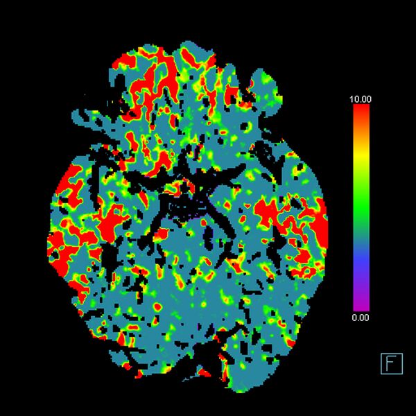
Original file (2,324 × 2,324 pixels, file size: 1.15 MB, MIME type: image/jpeg)
Summary:
- Radiopaedia case ID: 72038
- Image ID: 51710446
- Image stack position: 12/31
- Aux modality: MTT
- Study description: CT perfusion
- Study findings: The old right ACA territory infarct demonstrates markedly decreased CBV and CBF as expected. Similar changes in the left temporal pole. Decreased MTT, Tmax and TTD in the right cerebral hemisphere affect the ACA and MCA territories and spares the occipital lobe (PCA territory). No corresponding CBF or CBV abnormality on the right to suggest core infarct.
- Modality: CT
- System: Interventional
- Findings: Hypodense gliosis in the right ACA territory in keeping with prior infarct. Similar post-ischemic gliosis in the left temporal lobe. No new area of grey-white matter differentiation loss. No insular ribbon sign. No dense vessel sign evident on the thin data set. No hemorrhage, surface collection, mass effect or midline shift. Ex vacuo dilatation of the right lateral ventricle. Prominent CSF in the right CP angle with mild mass effect on the right cerebellar hemisphere is an arachnoid cyst which is stable.
- Published: 5th Nov 2019
- Source: https://radiopaedia.org/cases/acute-ica-ischaemic-penumbra-due-to-high-grade-cca-stenosis-ct-perfusion
- Author: Craig Hacking
- Permission: http://creativecommons.org/licenses/by-nc-sa/3.0/
Licensing:
Attribution-NonCommercial-ShareAlike 3.0 Unported (CC BY-NC-SA 3.0)
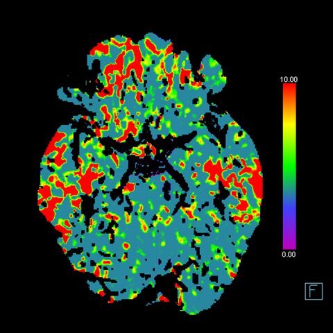
File history
Click on a date/time to view the file as it appeared at that time.
| Date/Time | Thumbnail | Dimensions | User | Comment | |
|---|---|---|---|---|---|
| current | 10:36, 11 April 2021 | | 2,324 × 2,324 (1.15 MB) | Fæ (talk | contribs) | Radiopaedia project rID:72038 (batch #872-13 B12) |
You cannot overwrite this file.
File usage
There are no pages that use this file.
_(Radiopaedia_72038-82531_MTT_12).jpg&oldid=114277)