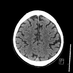
File:Acute P1 occlusion with PCA ischemia penumbra (CT perfusion) (Radiopaedia 72084-82586 Axial non-contrast 34).jpg
Jump to navigation
Jump to search

Size of this preview: 600 × 600 pixels. Other resolutions: 240 × 240 pixels | 480 × 480 pixels | 768 × 768 pixels | 1,024 × 1,024 pixels | 2,324 × 2,324 pixels.
Original file (2,324 × 2,324 pixels, file size: 654 KB, MIME type: image/jpeg)
Summary:
| Description |
|
| Date | Published: 7th Nov 2019 |
| Source | https://radiopaedia.org/cases/acute-p1-occlusion-with-pca-ischaemia-penumbra-ct-perfusion |
| Author | Craig Hacking |
| Permission (Permission-reusing-text) |
http://creativecommons.org/licenses/by-nc-sa/3.0/ |
Licensing:
Attribution-NonCommercial-ShareAlike 3.0 Unported (CC BY-NC-SA 3.0)
File history
Click on a date/time to view the file as it appeared at that time.
| Date/Time | Thumbnail | Dimensions | User | Comment | |
|---|---|---|---|---|---|
| current | 17:22, 16 April 2021 | | 2,324 × 2,324 (654 KB) | Fæ (talk | contribs) | Radiopaedia project rID:72084 (batch #979-34 A34) |
You cannot overwrite this file.
File usage
There are no pages that use this file.
_(Radiopaedia_72084-82586_Axial_non-contrast_34).jpg&oldid=148205)