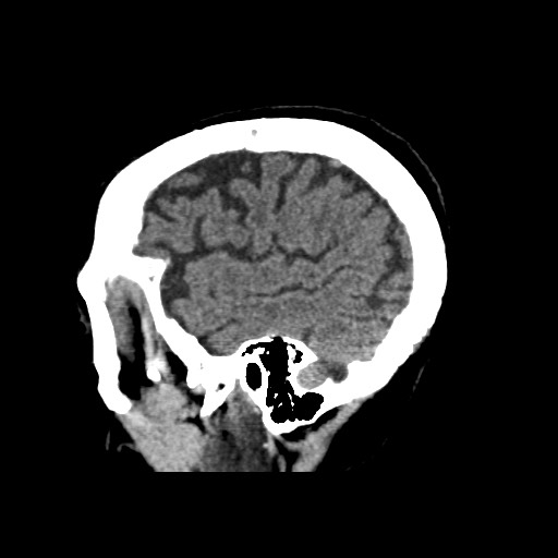
File:Acute PCA infarct (Radiopaedia 57048-63928 Sagittal non-contrast 13).jpg
Jump to navigation
Jump to search
Acute_PCA_infarct_(Radiopaedia_57048-63928_Sagittal_non-contrast_13).jpg (512 × 512 pixels, file size: 32 KB, MIME type: image/jpeg)
Summary:
| Description |
|
| Date | Published: 20th Feb 2018 |
| Source | https://radiopaedia.org/cases/acute-pca-infarct |
| Author | Andrew Dixon |
| Permission (Permission-reusing-text) |
http://creativecommons.org/licenses/by-nc-sa/3.0/ |
Licensing:
Attribution-NonCommercial-ShareAlike 3.0 Unported (CC BY-NC-SA 3.0)
File history
Click on a date/time to view the file as it appeared at that time.
| Date/Time | Thumbnail | Dimensions | User | Comment | |
|---|---|---|---|---|---|
| current | 23:37, 17 April 2021 |  | 512 × 512 (32 KB) | Fæ (talk | contribs) | Radiopaedia project rID:57048 (batch #1030-153 C13) |
You cannot overwrite this file.
File usage
The following page uses this file:
