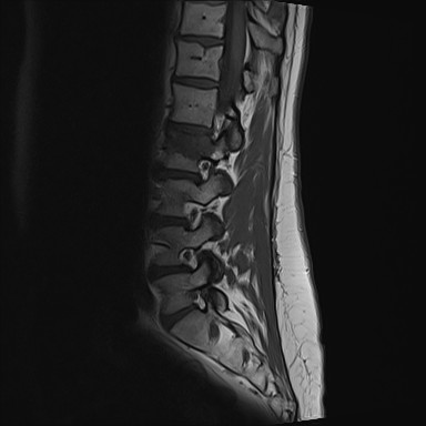
File:Acute Schmorl's node (Radiopaedia 83276-97685 Sagittal T1 17).jpg
Jump to navigation
Jump to search
Acute_Schmorl's_node_(Radiopaedia_83276-97685_Sagittal_T1_17).jpg (384 × 384 pixels, file size: 19 KB, MIME type: image/jpeg)
Summary:
| Description |
|
| Date | Published: 28th Jan 2021 |
| Source | https://radiopaedia.org/cases/acute-schmorls-node-3 |
| Author | Henry Knipe |
| Permission (Permission-reusing-text) |
http://creativecommons.org/licenses/by-nc-sa/3.0/ |
Licensing:
Attribution-NonCommercial-ShareAlike 3.0 Unported (CC BY-NC-SA 3.0)
File history
Click on a date/time to view the file as it appeared at that time.
| Date/Time | Thumbnail | Dimensions | User | Comment | |
|---|---|---|---|---|---|
| current | 10:01, 19 April 2021 |  | 384 × 384 (19 KB) | Fæ (talk | contribs) | Radiopaedia project rID:83276 (batch #1099-17 A17) |
You cannot overwrite this file.
File usage
There are no pages that use this file.
