File:Acute and chronic vertebral compression fractures (Radiopaedia 31608-32494 Coronal bone window 40).jpg
Jump to navigation
Jump to search

Size of this preview: 600 × 600 pixels. Other resolutions: 240 × 240 pixels | 480 × 480 pixels | 768 × 768 pixels | 1,024 × 1,024 pixels | 1,512 × 1,512 pixels.
Original file (1,512 × 1,512 pixels, file size: 271 KB, MIME type: image/jpeg)
Summary:
- Radiopaedia case ID: 31608
- Image ID: 8714147
- Image stack position: 40/43
- Plane projection: Coronal
- Aux modality: bone window
- Study description: Thoracic Spine CT
- Modality: CT
- System: Trauma
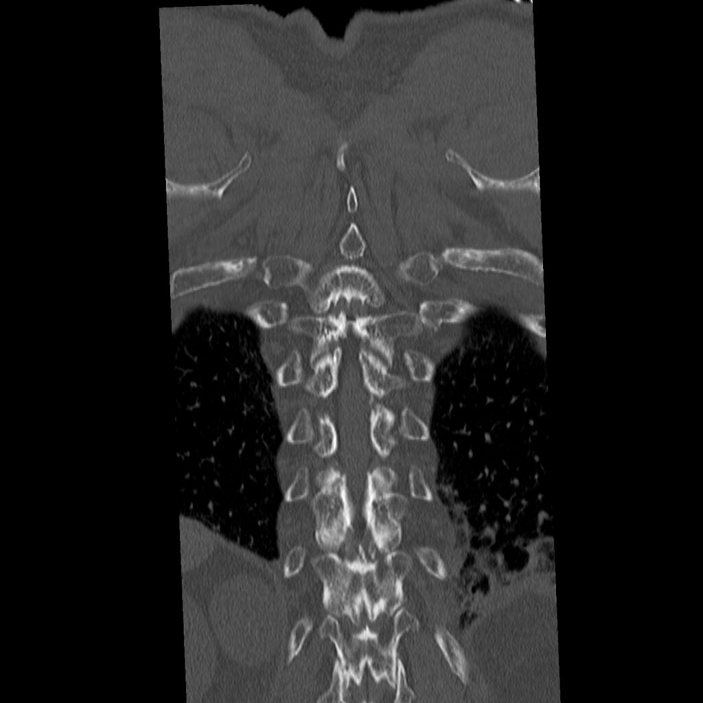
- Findings: 13 paired ribs. The first rib bearing vertebra is taken as T1. Buckling of the superior portion of the T6 and T10 vertebral bodies consistent with acute fractures. Note the angulation of the anterior and lateral cortex at these two levels and subtle compressive sclerosis in the medullary cavity where the normal trabeculae have been impacted. Old vertebral body compression fractures at T9 and L1. Note the remodeled cortex and endplate at these levels without any sharp buckling of the cortex.
- Published: 18th Oct 2014
- Source: https://radiopaedia.org/cases/acute-and-chronic-vertebral-compression-fractures
- Author: Andrew Dixon
- Permission: http://creativecommons.org/licenses/by-nc-sa/3.0/
Licensing:
Attribution-NonCommercial-ShareAlike 3.0 Unported (CC BY-NC-SA 3.0)
File history
Click on a date/time to view the file as it appeared at that time.
| Date/Time | Thumbnail | Dimensions | User | Comment | |
|---|---|---|---|---|---|
| current | 03:41, 4 April 2021 | | 1,512 × 1,512 (271 KB) | Fæ (talk | contribs) | Radiopaedia project rID:31608 (batch #650-70 B40) |
You cannot overwrite this file.
File usage
There are no pages that use this file.
.jpg&oldid=114423)