File:Acute anteroseptal myocardial infarction (Radiopaedia 68409-77945 Short axis (basal) Perfusion 31).jpg
Jump to navigation
Jump to search

Size of this preview: 552 × 599 pixels. Other resolutions: 221 × 240 pixels | 442 × 480 pixels | 707 × 768 pixels | 943 × 1,024 pixels | 1,334 × 1,448 pixels.
Original file (1,334 × 1,448 pixels, file size: 153 KB, MIME type: image/jpeg)
Summary:
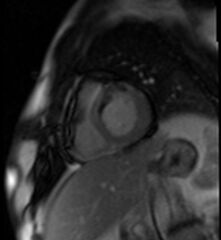
- Radiopaedia case ID: 68409
- Image ID: 49019546
- Image stack position: 31/50
- Plane projection: Short axis (basal)
- Aux modality: Perfusion
- Study description: Perfusion and viability module (Gadolinium first pass and LGE)
- Study findings: First pass or perfusion images: subendocardial perfusion defect (absence of signal, dark or black) in LAD territory. Late gadolinium enhancement: depicts transmural infarction (transmural hyperintensity, bright or white), also shows "no reflow" phenomenon (endocardial rim of absence of signal even in LGE images). Revascularization has a poor outcome in transmural infraction (considered non viable tissue) and the presence of "no reflow" phenomenon has poor prognosis.
- Modality: MRI
- System: Cardiac
- Findings: Anterior and septal akynesia. Pericardial effusion.
- Published: 29th May 2019
- Source: https://radiopaedia.org/cases/acute-anteroseptal-myocardial-infarction
- Author: David Cuevas
- Permission: http://creativecommons.org/licenses/by-nc-sa/3.0/
Licensing:
Attribution-NonCommercial-ShareAlike 3.0 Unported (CC BY-NC-SA 3.0)
File history
Click on a date/time to view the file as it appeared at that time.
| Date/Time | Thumbnail | Dimensions | User | Comment | |
|---|---|---|---|---|---|
| current | 04:06, 4 April 2021 | | 1,334 × 1,448 (153 KB) | Fæ (talk | contribs) | Radiopaedia project rID:68409 (batch #651-31 A31) |
You cannot overwrite this file.
File usage
There are no pages that use this file.
_Perfusion_31).jpg&oldid=87486)