File:Acute anteroseptal myocardial infarction (Radiopaedia 68409-77947 Short axis T2 3).jpg
Jump to navigation
Jump to search

Size of this preview: 600 × 600 pixels. Other resolutions: 240 × 240 pixels | 480 × 480 pixels | 768 × 768 pixels | 1,024 × 1,024 pixels | 1,448 × 1,448 pixels.
Original file (1,448 × 1,448 pixels, file size: 293 KB, MIME type: image/jpeg)
Summary:
- Radiopaedia case ID: 68409
- Image ID: 49019154
- Image stack position: 3/3
- Plane projection: Short axis
- Aux modality: T2
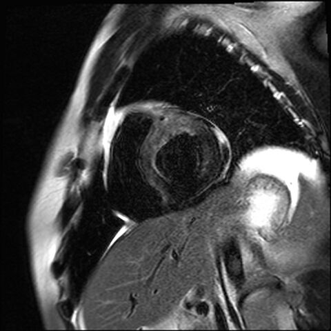
- Study description: Tissue characterization (no contrast)
- Study findings: T2 weighted images show extensive septal and anterior edema. Gradient echo image depicts a dark anteroseptal and anterior wall rim consistent with myocardial hemorrhage. DWI, optional/experimental use in myocardial infraction, shows both findings.
- Modality: MRI
- System: Cardiac
- Findings: Anterior and septal akynesia. Pericardial effusion.
- Published: 29th May 2019
- Source: https://radiopaedia.org/cases/acute-anteroseptal-myocardial-infarction
- Author: David Cuevas
- Permission: http://creativecommons.org/licenses/by-nc-sa/3.0/
Licensing:
Attribution-NonCommercial-ShareAlike 3.0 Unported (CC BY-NC-SA 3.0)
File history
Click on a date/time to view the file as it appeared at that time.
| Date/Time | Thumbnail | Dimensions | User | Comment | |
|---|---|---|---|---|---|
| current | 03:59, 4 April 2021 | | 1,448 × 1,448 (293 KB) | Fæ (talk | contribs) | Radiopaedia project rID:68409 (batch #651-3 A3) |
You cannot overwrite this file.
File usage
There are no pages that use this file.
.jpg&oldid=114665)