File:Acute aortic dissection - Stanford type A (Radiopaedia 40661-43285 Axial C+ portal venous phase 63).jpg
Jump to navigation
Jump to search
Size of this preview: 657 × 599 pixels. Other resolutions: 263 × 240 pixels | 526 × 480 pixels | 662 × 604 pixels.
Original file (662 × 604 pixels, file size: 125 KB, MIME type: image/jpeg)
Summary:
- Radiopaedia case ID: 40661
- Image ID: 17001776
- Image stack position: 63/120
- Plane projection: Axial
- Aux modality: C+ portal venous phase
- Modality: CT
- System: Vascular
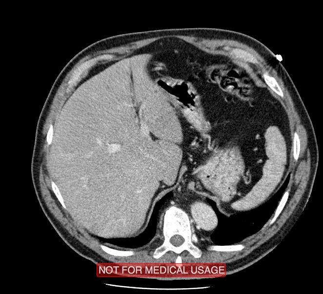
- Findings: Computed tomography angiography of whole aorta was performed. Intimal flap was identified from aortic valvolar plane to external iliac arteries, with true lumen and false lumen equally opacified by contrast medium in arterial phase. Small hematoma surrounding thoracic ascending aorta was seen. Epiaortic arteries wasn't involed; celiac trunk, superior mesentery artery originate from true lumen. Right renal artery originates from true lumen and it is involved by intimal flap in its proximal tract, with normal renal perfusion. Left renal artery originates from false lumen with left renal hypoperfusion.
- Published: 15th Nov 2015
- Source: https://radiopaedia.org/cases/acute-aortic-dissection-stanford-type-a-2
- Author: Giovanni Rinaldi
- Permission: http://creativecommons.org/licenses/by-nc-sa/3.0/
Licensing:
Attribution-NonCommercial-ShareAlike 3.0 Unported (CC BY-NC-SA 3.0)
File history
Click on a date/time to view the file as it appeared at that time.
| Date/Time | Thumbnail | Dimensions | User | Comment | |
|---|---|---|---|---|---|
| current | 05:37, 4 April 2021 | | 662 × 604 (125 KB) | Fæ (talk | contribs) | Radiopaedia project rID:40661 (batch #652-263 C63) |
You cannot overwrite this file.
File usage
The following page uses this file:
.jpg&oldid=88356)