File:Acute appendicitis (Radiopaedia 22892-22918 Coronal C+ portal venous phase 54).png
Jump to navigation
Jump to search

Size of this preview: 394 × 599 pixels. Other resolutions: 158 × 240 pixels | 442 × 672 pixels.
Original file (442 × 672 pixels, file size: 244 KB, MIME type: image/png)
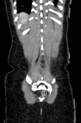
Summary:
- Radiopaedia case ID: 22892
- Image ID: 3333916
- Image stack position: 54/60
- Plane projection: Coronal
- Aux modality: C+ portal venous phase
- Study description: CT abdomen and pelvis
- Modality: CT
- System: Gastrointestinal
- Findings: Cecal appendix in the right lower quadrant is increased in diameter, reaching 10 mm, with wall thickening and mucosal hyper-enhancement associated with increased periappendiceal fat density and cecal pole edema. No collections, free fluid or pneumoperitoneum are identified.
- Published: 1st May 2013
- Source: https://radiopaedia.org/cases/acute-appendicitis-12
- Author: David Cuete
- Permission: http://creativecommons.org/licenses/by-nc-sa/3.0/
Licensing:
Attribution-NonCommercial-ShareAlike 3.0 Unported (CC BY-NC-SA 3.0)
File history
Click on a date/time to view the file as it appeared at that time.
| Date/Time | Thumbnail | Dimensions | User | Comment | |
|---|---|---|---|---|---|
| current | 10:10, 4 April 2021 | | 442 × 672 (244 KB) | Fæ (talk | contribs) | Radiopaedia project rID:22892 (batch #669-139 B54) |
You cannot overwrite this file.
File usage
The following page uses this file:
.png&oldid=91399)