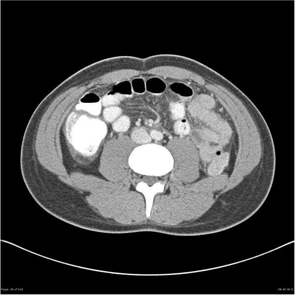
File:Acute appendicitis (Radiopaedia 25364-25615 B 36).jpg
Jump to navigation
Jump to search

Size of this preview: 600 × 600 pixels. Other resolutions: 240 × 240 pixels | 480 × 480 pixels | 768 × 768 pixels | 1,024 × 1,024 pixels | 1,365 × 1,365 pixels.
Original file (1,365 × 1,365 pixels, file size: 407 KB, MIME type: image/jpeg)
Summary:
- Radiopaedia case ID: 25364
- Image ID: 4878480
- Image stack position: 36/76
- Aux modality: C+ portal venous phase
- Modality: CT
- System: Gastrointestinal
- Findings: The appendix, located near the midline, is thickened / edematous, measuring 10 mm in maximal diameter. Adjacent peri appendiceal fat stranding as well is moderate volume of free intra-abdominal fluid is demonstrated. No drainable collection. Oral contrast passes through to the transverse colon, no evidence of bowel obstruction. No free gas. The liver, spleen, pancreas, kidneys and adrenals are within normal limits. Several small reactive mesenteric lymph nodes are demonstrated. Dependent atelectasis in the lung bases and small bilateral pleural effusions. The bones are within normal limits.
- Published: 22nd Oct 2013
- Source: https://radiopaedia.org/cases/acute-appendicitis-16
- Author: Frank Gaillard
- Permission: http://creativecommons.org/licenses/by-nc-sa/3.0/
Licensing:
Attribution-NonCommercial-ShareAlike 3.0 Unported (CC BY-NC-SA 3.0)
File history
Click on a date/time to view the file as it appeared at that time.
| Date/Time | Thumbnail | Dimensions | User | Comment | |
|---|---|---|---|---|---|
| current | 12:43, 4 April 2021 |  | 1,365 × 1,365 (407 KB) | Fæ (talk | contribs) | Radiopaedia project rID:25364 (batch #676-62 B36) |
You cannot overwrite this file.
File usage
The following page uses this file: