File:Acute appendicitis (Radiopaedia 27049-27227 Axial C+ portal venous phase 36).jpg
Jump to navigation
Jump to search
Size of this preview: 600 × 600 pixels. Other resolutions: 240 × 240 pixels | 613 × 613 pixels.
Original file (613 × 613 pixels, file size: 78 KB, MIME type: image/jpeg)
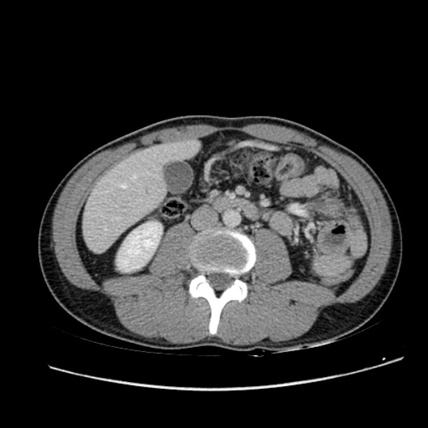
Summary:
- Radiopaedia case ID: 27049
- Image ID: 5640392
- Image stack position: 36/80
- Plane projection: Axial
- Aux modality: C+ portal venous phase
- Study description: CT abdomen and pelvis
- Modality: CT
- System: Gastrointestinal
- Findings: There is a distended appendix with an enhancing wall and two appendicoliths. Stranding is seen in the periappendiceal fat. No abnormal periappendiceal fluid collection or free gas can be seen. Few mesenteric lymph nodes in the vicinity of the inflamed appendix. Incidental liver cyst in segment VIII.
- Published: 19th Jan 2014
- Source: https://radiopaedia.org/cases/acute-appendicitis-18
- Author: David Cuete
- Permission: http://creativecommons.org/licenses/by-nc-sa/3.0/
Licensing:
Attribution-NonCommercial-ShareAlike 3.0 Unported (CC BY-NC-SA 3.0)
File history
Click on a date/time to view the file as it appeared at that time.
| Date/Time | Thumbnail | Dimensions | User | Comment | |
|---|---|---|---|---|---|
| current | 19:06, 4 April 2021 | | 613 × 613 (78 KB) | Fæ (talk | contribs) | Radiopaedia project rID:27049 (batch #698-36 A36) |
You cannot overwrite this file.
File usage
The following page uses this file:
.jpg&oldid=97180)