File:Acute appendicitis (Radiopaedia 30297-30934 Axial C+ portal venous phase 83).jpg
Jump to navigation
Jump to search
No higher resolution available.
Acute_appendicitis_(Radiopaedia_30297-30934_Axial_C+_portal_venous_phase_83).jpg (512 × 512 pixels, file size: 31 KB, MIME type: image/jpeg)
Summary:
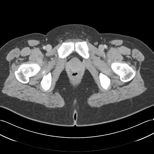
- Radiopaedia case ID: 30297
- Image ID: 7844182
- Image stack position: 83/87
- Plane projection: Axial
- Aux modality: C+ portal venous phase
- Modality: CT
- System: Gastrointestinal
- Findings: Moderate fat stranding surrounds the dilated (15 mm) fluid filled appendix arising from the lateral aspect of the cecal pole and extending inferiorly and anteriorly, measuring approximately 7 cm in total length. No collection or evidence of perforation identified. No free fluid or pneumoperitoneum. The remainder of the bowel and solid organs appear normal. Multiple prominent subcentimeter mesenteric and retroperitoneal lymph nodes. Conclusion Appendicitis without evidence of perforation. The multiple enlarged draining lymph nodes are probably reactive.
- Published: 3rd Aug 2014
- Source: https://radiopaedia.org/cases/acute-appendicitis-22
- Author: RMH Core Conditions
- Permission: http://creativecommons.org/licenses/by-nc-sa/3.0/
Licensing:
Attribution-NonCommercial-ShareAlike 3.0 Unported (CC BY-NC-SA 3.0)
File history
Click on a date/time to view the file as it appeared at that time.
| Date/Time | Thumbnail | Dimensions | User | Comment | |
|---|---|---|---|---|---|
| current | 13:18, 4 April 2021 | | 512 × 512 (31 KB) | Fæ (talk | contribs) | Radiopaedia project rID:30297 (batch #679-83 A83) |
You cannot overwrite this file.
File usage
The following page uses this file:
.jpg&oldid=93734)