File:Acute appendicitis (Radiopaedia 40656).jpg
Jump to navigation
Jump to search

Size of this preview: 542 × 599 pixels. Other resolutions: 217 × 240 pixels | 434 × 480 pixels | 681 × 753 pixels.
Original file (681 × 753 pixels, file size: 94 KB, MIME type: image/jpeg)
Summary:
- Radiopaedia case ID: 40656
- Image ID: 16964089
- Study description: CT Abdomen and pelvis
- Modality: CT
- System: Gastrointestinal
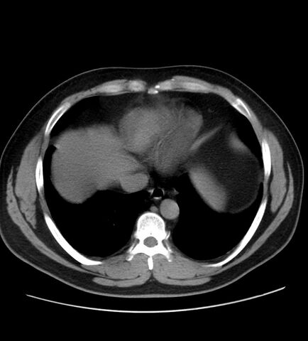
- Findings: The appendix s diffusely enlarged (measuring 2,0 cm in diameter), with thickened walls, and surrounded by periappendiceal inflammation, including stranding of the adjacent fat and minimal extraluminal fluid. Two small calcifications within the appendix base may correspond to appendicolith. There is also a diffuse hepatic steatosis. Remainder exam is unremarkable.
- Published: 29th Oct 2015
- Source: https://radiopaedia.org/cases/acute-appendicitis-32
- Author: Bruno Di Muzio
- Permission: http://creativecommons.org/licenses/by-nc-sa/3.0/
Licensing:
Attribution-NonCommercial-ShareAlike 3.0 Unported (CC BY-NC-SA 3.0)
File history
Click on a date/time to view the file as it appeared at that time.
| Date/Time | Thumbnail | Dimensions | User | Comment | |
|---|---|---|---|---|---|
| current | 10:54, 4 April 2021 | | 681 × 753 (94 KB) | Fæ (talk | contribs) | Radiopaedia project rID:40656 (batch #672) |
You cannot overwrite this file.
File usage
The following page uses this file:
.jpg&oldid=8854272)