File:Acute appendicitis (Radiopaedia 51979).jpg
Jump to navigation
Jump to search

Size of this preview: 738 × 600 pixels. Other resolutions: 295 × 240 pixels | 591 × 480 pixels | 945 × 768 pixels | 1,024 × 832 pixels.
Original file (1,024 × 832 pixels, file size: 94 KB, MIME type: image/jpeg)
Summary:
- Radiopaedia case ID: 51979
- Image ID: 29171274
- Modality: Ultrasound
- System: Gastrointestinal
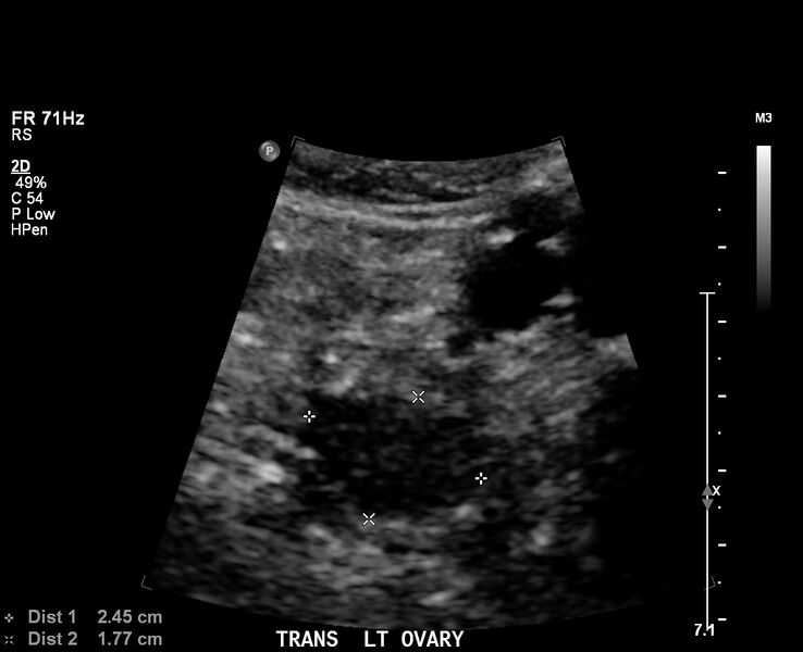
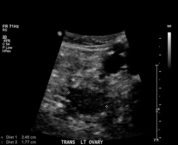
- Findings: Aperistaltic, non-compressible, dilated vermiform appendix is seen in the right lower fossa measuring 9 mm in diameter, with hyperemia and surrounding hyperechoic fat. 'Target" appearance of the appendix on axial plane. Tenderness on ultrasound probe palpation of the vermiform appendix. Normal-sized and non-tender bilateral ovaries on trans-abdominal scanning
- Published: 16th Mar 2017
- Source: https://radiopaedia.org/cases/acute-appendicitis-57
- Author: Matthew Lukies
- Permission: http://creativecommons.org/licenses/by-nc-sa/3.0/
Licensing:
Attribution-NonCommercial-ShareAlike 3.0 Unported (CC BY-NC-SA 3.0)
File history
Click on a date/time to view the file as it appeared at that time.
| Date/Time | Thumbnail | Dimensions | User | Comment | |
|---|---|---|---|---|---|
| current | 20:35, 4 April 2021 | | 1,024 × 832 (94 KB) | Fæ (talk | contribs) | Radiopaedia project rID:51979 (batch #702) |
You cannot overwrite this file.
File usage
The following page uses this file:
.jpg&oldid=8854263)