File:Acute appendicitis (Radiopaedia 53100-74098 Surgical specimen 1).jpeg
Jump to navigation
Jump to search
Size of this preview: 337 × 599 pixels. Other resolutions: 135 × 240 pixels | 270 × 480 pixels | 720 × 1,280 pixels.
Original file (720 × 1,280 pixels, file size: 85 KB, MIME type: image/jpeg)
Summary:
- Radiopaedia case ID: 53100
- Image ID: 44896165
- Plane projection: Surgical specimen
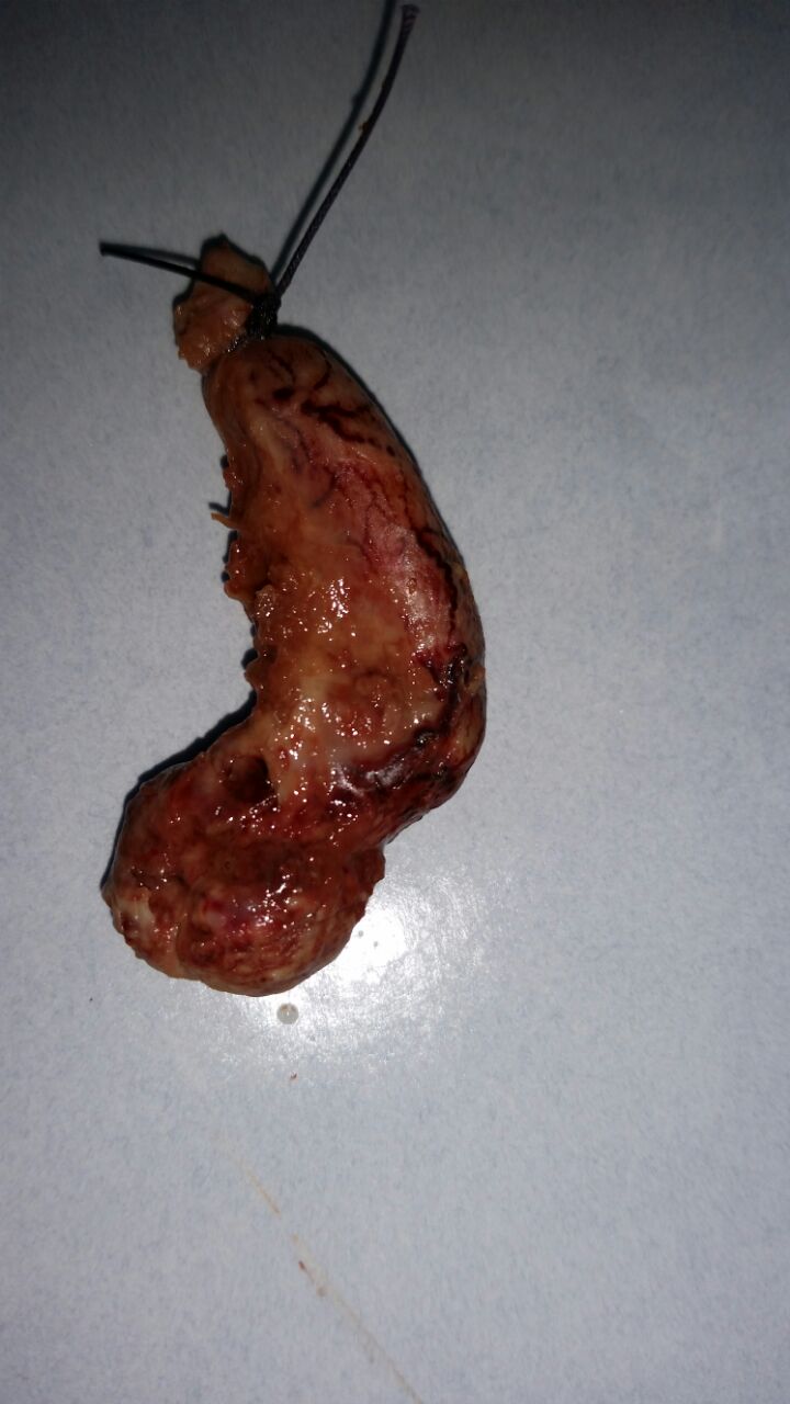
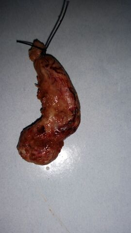
- Study findings: Inflamed appendix with gangrenous areas and focal perforation.
- Modality: Photo
- System: Gastrointestinal
- Findings: Thick-walled markedly enlarged appendix with heterogeneous content. Increased wall vascularity on color Doppler examination. An adjacent small amount of turbid fluid is seen.
- Published: 23rd Dec 2018
- Source: https://radiopaedia.org/cases/acute-appendicitis-43
- Author: Sameh Saied Ali
- Permission: http://creativecommons.org/licenses/by-nc-sa/3.0/
Licensing:
Attribution-NonCommercial-ShareAlike 3.0 Unported (CC BY-NC-SA 3.0)
File history
Click on a date/time to view the file as it appeared at that time.
| Date/Time | Thumbnail | Dimensions | User | Comment | |
|---|---|---|---|---|---|
| current | 08:04, 4 April 2021 | | 720 × 1,280 (85 KB) | Fæ (talk | contribs) | Radiopaedia project rID:53100 (batch #659-1 A1) |
You cannot overwrite this file.
File usage
There are no pages that use this file.
.jpeg&oldid=89963)