File:Acute appendicitis (Radiopaedia 62608-70901 Axial C+ portal venous phase 22).jpg
Jump to navigation
Jump to search
No higher resolution available.
Acute_appendicitis_(Radiopaedia_62608-70901_Axial_C+_portal_venous_phase_22).jpg (512 × 512 pixels, file size: 94 KB, MIME type: image/jpeg)
Summary:
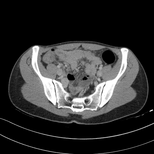
- Radiopaedia case ID: 62608
- Image ID: 42013030
- Image stack position: 22/75
- Plane projection: Axial
- Aux modality: C+ portal venous phase
- Modality: CT
- System: Gastrointestinal
- Findings: The appendix is thickened (see key image), and its wall shows contrast enhancement especially in the distal part. The appendix is arching down deep into the pelvis, explaining why visualization with ultrasound was likely unsuccessful. Surrounding fat stranding an low density periappendicular and pelvic free fluid (key image 2) is observed. A relative increase of the size of the mesenteric lymph nodes is visible in the right lower quadrant. Altogether the findings are in line with the diagnosis of acute appendicitis. The inflamed appendix was successfuly removed laparoscopically.
- Published: 24th Aug 2018
- Source: https://radiopaedia.org/cases/acute-appendicitis-45
- Author: Balint Botz
- Permission: http://creativecommons.org/licenses/by-nc-sa/3.0/
Licensing:
Attribution-NonCommercial-ShareAlike 3.0 Unported (CC BY-NC-SA 3.0)
File history
Click on a date/time to view the file as it appeared at that time.
| Date/Time | Thumbnail | Dimensions | User | Comment | |
|---|---|---|---|---|---|
| current | 08:31, 4 April 2021 | | 512 × 512 (94 KB) | Fæ (talk | contribs) | Radiopaedia project rID:62608 (batch #665-22 A22) |
You cannot overwrite this file.
File usage
The following page uses this file:
.jpg&oldid=90284)