File:Acute appendicitis (Radiopaedia 77016-88960 Coronal C+ portal venous phase 17).jpg
Jump to navigation
Jump to search
No higher resolution available.
Acute_appendicitis_(Radiopaedia_77016-88960_Coronal_C+_portal_venous_phase_17).jpg (512 × 547 pixels, file size: 32 KB, MIME type: image/jpeg)
Summary:
- Radiopaedia case ID: 77016
- Image ID: 52533922
- Image stack position: 17/101
- Plane projection: Coronal
- Aux modality: C+ portal venous phase
- Modality: CT
- System: Gastrointestinal
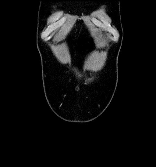
- Findings: The appendix is dilated to 24 mm. The wall is thickened and there is surrounding fat stranding, focal fluid and thickening of the adjacent parietal peritoneum. Two appendicoliths can be identified, one at the base of the appendix and the other at the tip. There is no abscess formation and no free intra-abdominal gas. The cecal wall is thickened. Secondary findings are a colonic diverticulosis.
- Published: 4th May 2020
- Source: https://radiopaedia.org/cases/acute-appendicitis-67
- Author: Muhammad Taha Hagar
- Permission: http://creativecommons.org/licenses/by-nc-sa/3.0/
Licensing:
Attribution-NonCommercial-ShareAlike 3.0 Unported (CC BY-NC-SA 3.0)
File history
Click on a date/time to view the file as it appeared at that time.
| Date/Time | Thumbnail | Dimensions | User | Comment | |
|---|---|---|---|---|---|
| current | 16:18, 4 April 2021 | | 512 × 547 (32 KB) | Fæ (talk | contribs) | Radiopaedia project rID:77016 (batch #686-181 B17) |
You cannot overwrite this file.
File usage
The following page uses this file:
.jpg&oldid=95637)