File:Acute appendicitis (Radiopaedia 85193-100745 D 4).jpg
Jump to navigation
Jump to search
No higher resolution available.
Acute_appendicitis_(Radiopaedia_85193-100745_D_4).jpg (640 × 441 pixels, file size: 111 KB, MIME type: image/jpeg)
Summary:
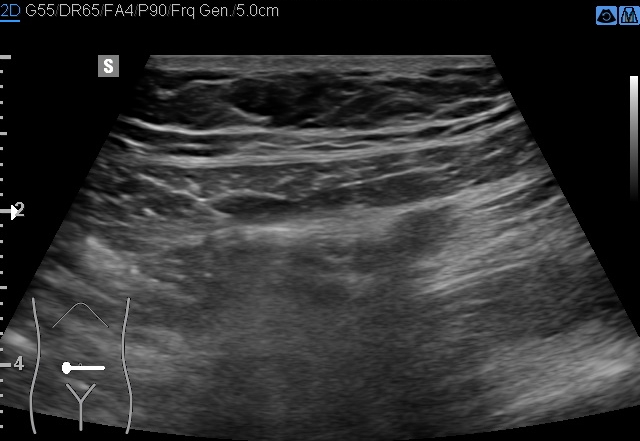
- Radiopaedia case ID: 85193
- Image ID: 54083657
- Image stack position: 4/225
- Plane projection: Oblique
- Modality: Ultrasound
- System: Gastrointestinal
- Findings: The vermiform appendix is coursing superficially and medially, well-visualized even with the curvilinear probe. Note marked thickening (13-15 mm) and wall irregularity. Painful and rigid upon direct compression. Hypervascularity with mesoappendiceal vasa recta engorgement. Periappendicular fat stranding and a small amount of fluid are also present.
- Published: 17th Dec 2020
- Source: https://radiopaedia.org/cases/acute-appendicitis-79
- Author: Balint Botz
- Permission: http://creativecommons.org/licenses/by-nc-sa/3.0/
Licensing:
Attribution-NonCommercial-ShareAlike 3.0 Unported (CC BY-NC-SA 3.0)
File history
Click on a date/time to view the file as it appeared at that time.
| Date/Time | Thumbnail | Dimensions | User | Comment | |
|---|---|---|---|---|---|
| current | 14:34, 4 April 2021 | | 640 × 441 (111 KB) | Fæ (talk | contribs) | Radiopaedia project rID:85193 (batch #681-363 D4) |
You cannot overwrite this file.
File usage
The following page uses this file:
.jpg&oldid=94596)