File:Acute appendicitis arising from a malrotated cecum (Radiopaedia 19970-19997 Coronal C+ portal venous phase 1).jpg
Jump to navigation
Jump to search
Size of this preview: 507 × 599 pixels. Other resolutions: 203 × 240 pixels | 512 × 605 pixels.
Original file (512 × 605 pixels, file size: 165 KB, MIME type: image/jpeg)
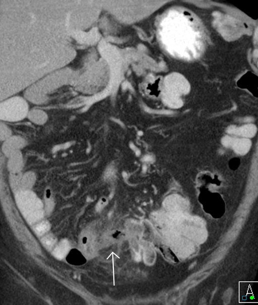
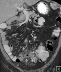
Summary:
- Radiopaedia case ID: 19970
- Image ID: 2480886
- Plane projection: Coronal
- Aux modality: C+ portal venous phase
- Modality: CT
- System: Gastrointestinal
- Findings: Findings typical of appendicitis with thickening of the wall and surrounding soft tissue swelling. Resultant inflammatory mass incorporating the terminal ileum with secondary mural thickening. Note the position of the ileo-cecal junction and appendix in the midline with colon to the left and small bowel to the right, as seen in malrotation.
- Published: 26th Oct 2012
- Source: https://radiopaedia.org/cases/acute-appendicitis-arising-from-a-malrotated-caecum
- Author: Chris O'Donnell
- Permission: http://creativecommons.org/licenses/by-nc-sa/3.0/
Licensing:
Attribution-NonCommercial-ShareAlike 3.0 Unported (CC BY-NC-SA 3.0)
File history
Click on a date/time to view the file as it appeared at that time.
| Date/Time | Thumbnail | Dimensions | User | Comment | |
|---|---|---|---|---|---|
| current | 23:50, 4 April 2021 | | 512 × 605 (165 KB) | Fæ (talk | contribs) | Radiopaedia project rID:19970 (batch #707-46 C1) |
You cannot overwrite this file.
File usage
There are no pages that use this file.
.jpg&oldid=100601)