File:Acute appendicitis complicated by ovarian vein thrombophlebitis (Radiopaedia 16172-15851 Axial C+ portal venous phase 48).jpg
Jump to navigation
Jump to search
No higher resolution available.
Acute_appendicitis_complicated_by_ovarian_vein_thrombophlebitis_(Radiopaedia_16172-15851_Axial_C+_portal_venous_phase_48).jpg (512 × 512 pixels, file size: 131 KB, MIME type: image/jpeg)
Summary:
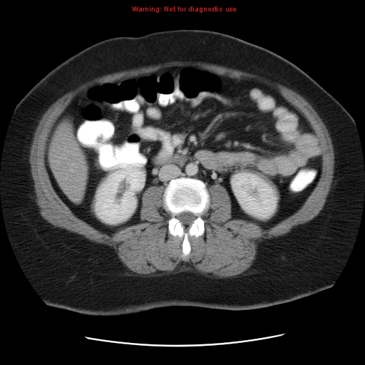
- Radiopaedia case ID: 16172
- Image ID: 1591754
- Image stack position: 48/96
- Plane projection: Axial
- Aux modality: C+ portal venous phase
- Modality: CT
- System: Gastrointestinal
- Findings: Dilated appendix with significant surrounding stranding. However, no drainable collection or extraluminal gas can be seen. The right ovarian vein is distended and has a thick enhancing wall, along with hypodensity in the lumen extending to the middle portion of the vein (the lumen is not enhancing vs the contralateral left ovarian vein). The changes of the right ovarian vein are better appreciated on the coronal images. No abdominal free fluid. No retroperitoneal or pelvic enlarged lymph nodes. Interpretation: Features of acute appendicitis complicated by ovarian vein thrombosis.
- Published: 26th Dec 2011
- Source: https://radiopaedia.org/cases/acute-appendicitis-complicated-by-ovarian-vein-thrombophlebitis
- Author: Hani Makky Al Salam
- Permission: http://creativecommons.org/licenses/by-nc-sa/3.0/
Licensing:
Attribution-NonCommercial-ShareAlike 3.0 Unported (CC BY-NC-SA 3.0)
File history
Click on a date/time to view the file as it appeared at that time.
| Date/Time | Thumbnail | Dimensions | User | Comment | |
|---|---|---|---|---|---|
| current | 23:59, 4 April 2021 | | 512 × 512 (131 KB) | Fæ (talk | contribs) | Radiopaedia project rID:16172 (batch #708-48 A48) |
You cannot overwrite this file.
File usage
The following page uses this file:
.jpg&oldid=100930)