File:Acute appendicitis with appendicoliths (Radiopaedia 50186-55532 Coronal C+ portal venous phase 45).png
Jump to navigation
Jump to search
Size of this preview: 501 × 599 pixels. Other resolutions: 201 × 240 pixels | 512 × 612 pixels.
Original file (512 × 612 pixels, file size: 118 KB, MIME type: image/png)
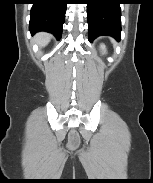
Summary:
- Radiopaedia case ID: 50186
- Image ID: 27226128
- Image stack position: 45/57
- Plane projection: Coronal
- Aux modality: C+ portal venous phase
- Study findings: The appendix is dilated with hyperenhancing walls with surrounding fat stranding. Two appendicoliths are seen at the base and ostia of the appendix. Small volume free fluid. No free gas.
- Modality: CT
- System: Gastrointestinal
- Findings: Non-specific bowel gas pattern with no convincing evidence of obstruction. RIF calcifications, probably fecoliths/appendicoliths. No free gas. Lung bases are clear.
- Published: 29th Dec 2016
- Source: https://radiopaedia.org/cases/acute-appendicitis-with-appendicoliths-1
- Author: Henry Knipe
- Permission: http://creativecommons.org/licenses/by-nc-sa/3.0/
Licensing:
Attribution-NonCommercial-ShareAlike 3.0 Unported (CC BY-NC-SA 3.0)
File history
Click on a date/time to view the file as it appeared at that time.
| Date/Time | Thumbnail | Dimensions | User | Comment | |
|---|---|---|---|---|---|
| current | 03:51, 5 April 2021 | | 512 × 612 (118 KB) | Fæ (talk | contribs) | Radiopaedia project rID:50186 (batch #723-139 B45) |
You cannot overwrite this file.
File usage
The following page uses this file:
.png&oldid=83427)