File:Acute bilateral pyelonephritis (Radiopaedia 37146-38881 Axial non-contrast 3).jpg
Jump to navigation
Jump to search
No higher resolution available.
Acute_bilateral_pyelonephritis_(Radiopaedia_37146-38881_Axial_non-contrast_3).jpg (512 × 512 pixels, file size: 44 KB, MIME type: image/jpeg)
Summary:
- Radiopaedia case ID: 37146
- Image ID: 13094779
- Image stack position: 3/67
- Plane projection: Axial
- Aux modality: non-contrast
- Modality: CT
- System: Urogenital
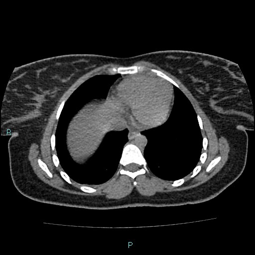
- Findings: Enlargement of both kidneys with heterogeneous parenchymal enhancement and multiple partially confluent hypoattenuating areas with blurred margins. There is a small cortical cyst at the lower pole of the left kidney. Marked fatty liver. Radiographer: TSRM Fabius Imola
- Published: 27th May 2015
- Source: https://radiopaedia.org/cases/acute-bilateral-pyelonephritis
- Author: Domenico Nicoletti
- Permission: http://creativecommons.org/licenses/by-nc-sa/3.0/
Licensing:
Attribution-NonCommercial-ShareAlike 3.0 Unported (CC BY-NC-SA 3.0)
File history
Click on a date/time to view the file as it appeared at that time.
| Date/Time | Thumbnail | Dimensions | User | Comment | |
|---|---|---|---|---|---|
| current | 15:24, 5 April 2021 | | 512 × 512 (44 KB) | Fæ (talk | contribs) | Radiopaedia project rID:37146 (batch #744-3 A3) |
You cannot overwrite this file.
File usage
The following page uses this file:
.jpg&oldid=70476)