File:Acute bleed from gastric fundus biopsy site (Radiopaedia 35201-36737 D 10).png
Jump to navigation
Jump to search
No higher resolution available.
Acute_bleed_from_gastric_fundus_biopsy_site_(Radiopaedia_35201-36737_D_10).png (512 × 571 pixels, file size: 138 KB, MIME type: image/png)
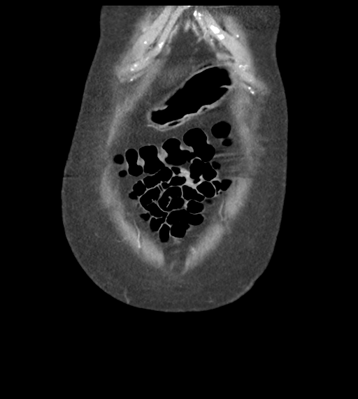
Summary:
- Radiopaedia case ID: 35201
- Image ID: 11869308
- Image stack position: 10/58
- Plane projection: Coronal
- Aux modality: C+ arterial phase
- Modality: CT
- System: Gastrointestinal
- Findings: Technique: non contrast, arterial and 30 sec delayed phases. IV contrast administered from the right leg. Large heterogenous density hematoma within the lumen of the dilated stomach. This is secondary to an acute arterial hemorrhage at the gastric fundus, seen on the arterial phase and pooling on the delayed phase. No other point of hemorrhage identified. IDC noted. Incidental calcified gallstones, no gallbladder wall thickening. The remaining intra-abdominal contents are unremarkable. Dependent consolidation in the lung bases represents atelectasis or aspiration. Small bilateral pleural effusions. Conclusion Arterial hemorrhage arising in the wall of the gastric fundus. This is at the site of recent endoscopic biopsy.
- Published: 28th Aug 2015
- Source: https://radiopaedia.org/cases/acute-bleed-from-gastric-fundus-biopsy-site
- Author: Craig Hacking
- Permission: http://creativecommons.org/licenses/by-nc-sa/3.0/
Licensing:
Attribution-NonCommercial-ShareAlike 3.0 Unported (CC BY-NC-SA 3.0)
File history
Click on a date/time to view the file as it appeared at that time.
| Date/Time | Thumbnail | Dimensions | User | Comment | |
|---|---|---|---|---|---|
| current | 19:21, 5 April 2021 | | 512 × 571 (138 KB) | Fæ (talk | contribs) | Radiopaedia project rID:35201 (batch #746-236 D10) |
You cannot overwrite this file.
File usage
The following page uses this file:
.png&oldid=71737)