File:Acute calcific periarthritis - wrist (Radiopaedia 76314).jpg
Jump to navigation
Jump to search

Size of this preview: 800 × 475 pixels. Other resolutions: 320 × 190 pixels | 640 × 380 pixels | 839 × 498 pixels.
Original file (839 × 498 pixels, file size: 50 KB, MIME type: image/jpeg)
Summary:
- Radiopaedia case ID: 76314
- Image ID: 52416560
- Modality: Ultrasound
- System: Musculoskeletal
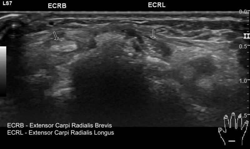
- Findings: At the site of swelling and pain pointed by the patient, there is an amorphous calcific deposit in the soft tissue involving the dorsum of the wrist. The calcification is located between tendons of 2nd extensor compartment and superficial to the carpal bone. There is a local hypervascularity. The extensor compartment tendons show normal echo pattern without tenosynovitis. The adjacent bone cortex does not show erosion.
- Published: 19th Apr 2020
- Source: https://radiopaedia.org/cases/acute-calcific-periarthritis-wrist-4
- Author: Maulik S Patel
- Permission: http://creativecommons.org/licenses/by-nc-sa/3.0/
Licensing:
Attribution-NonCommercial-ShareAlike 3.0 Unported (CC BY-NC-SA 3.0)
File history
Click on a date/time to view the file as it appeared at that time.
| Date/Time | Thumbnail | Dimensions | User | Comment | |
|---|---|---|---|---|---|
| current | 02:51, 6 April 2021 | | 839 × 498 (50 KB) | Fæ (talk | contribs) | Radiopaedia project rID:76314 (batch #752) |
You cannot overwrite this file.
File usage
The following page uses this file:
.jpg&oldid=8855591)