File:Acute calcular cholecystitis (Radiopaedia 87362-103682 Coronal 3D MRCP 50).jpg
Jump to navigation
Jump to search
No higher resolution available.
Acute_calcular_cholecystitis_(Radiopaedia_87362-103682_Coronal_3D_MRCP_50).jpg (512 × 512 pixels, file size: 45 KB, MIME type: image/jpeg)
Summary:
- Radiopaedia case ID: 87362
- Image ID: 54512765
- Image stack position: 50/85
- Plane projection: Coronal
- Aux modality: 3D MRCP
- Study description: MRCP on day 3
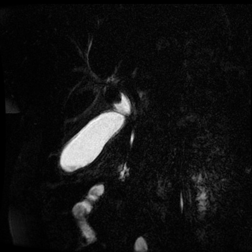
- Study findings: The gallbladder is distended and an impacted stone in its neck appearing as a rounded filling defect measuring about 2 cm is noted, with diffuse wall thickening and minimal pericholecystic inflammatory fatty stranding impressive of acute calcular cholecystitis. Normal caliber and outline of intrahepatic bile ducts, right/left common hepatic, cystic duct and common bile ducts. No evidence of free fluid or lymphadenopathy is detected. The bilateral minimal pleural reaction is noted. Conclusion:Features on MRI are compatible with acute cholecystitis with impacted stone at the neck of the gallbladder.
- Modality: MRI
- System: Hepatobiliary
- Findings: The gall bladder is distended with a large stone lodged in the Hartman pouch measuring about 2cm with echogenic biliary mud within and has diffuse wall thickening, but no pericholecystic fluid noted collections.
- Published: 4th Mar 2021
- Source: https://radiopaedia.org/cases/acute-calcular-cholecystitis-3
- Author: Safwat Mohammad Almoghazy
- Permission: http://creativecommons.org/licenses/by-nc-sa/3.0/
Licensing:
Attribution-NonCommercial-ShareAlike 3.0 Unported (CC BY-NC-SA 3.0)
File history
Click on a date/time to view the file as it appeared at that time.
| Date/Time | Thumbnail | Dimensions | User | Comment | |
|---|---|---|---|---|---|
| current | 04:04, 6 April 2021 | | 512 × 512 (45 KB) | Fæ (talk | contribs) | Radiopaedia project rID:87362 (batch #754-182 D50) |
You cannot overwrite this file.
File usage
The following page uses this file:
.jpg&oldid=74628)