File:Acute cerebral infarct (Radiopaedia 35604-37123 Axial non-contrast 41).jpg
Jump to navigation
Jump to search

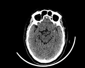
Size of this preview: 746 × 599 pixels. Other resolutions: 299 × 240 pixels | 598 × 480 pixels | 956 × 768 pixels | 1,275 × 1,024 pixels | 1,501 × 1,206 pixels.
Original file (1,501 × 1,206 pixels, file size: 227 KB, MIME type: image/jpeg)
Summary:
- Radiopaedia case ID: 35604
- Image ID: 12141309
- Image stack position: 41/83
- Plane projection: Axial
- Aux modality: non-contrast
- Modality: CT
- System: Central Nervous System
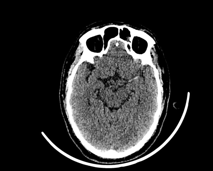
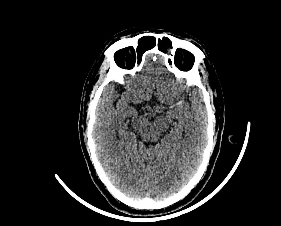
- Findings: The left middle cerebral artery appears hyperdense. There is an ill-defined hypodense region noted involving the left insular cortex with loss of grey and white matter interface. There loss of delineation of the basal ganglia suggestive of disappearing basal ganglia sign. Hyperdense MCA, loss of insular ribbon and disappearing basal ganglia signs are suggestive of acute cerebral infarct.
- Published: 13th Apr 2015
- Source: https://radiopaedia.org/cases/acute-cerebral-infarct-1
- Author: Prashant Mudgal
- Permission: http://creativecommons.org/licenses/by-nc-sa/3.0/
Licensing:
Attribution-NonCommercial-ShareAlike 3.0 Unported (CC BY-NC-SA 3.0)
File history
Click on a date/time to view the file as it appeared at that time.
| Date/Time | Thumbnail | Dimensions | User | Comment | |
|---|---|---|---|---|---|
| current | 18:15, 6 April 2021 | | 1,501 × 1,206 (227 KB) | Fæ (talk | contribs) | Radiopaedia project rID:35604 (batch #772-41 A41) |
You cannot overwrite this file.
File usage
The following page uses this file:
.jpg&oldid=79168)