File:Acute chest syndrome - sickle cell disease (Radiopaedia 42375-45499 Axial lung window 174).jpg
Jump to navigation
Jump to search

Size of this preview: 600 × 600 pixels. Other resolutions: 240 × 240 pixels | 480 × 480 pixels | 768 × 768 pixels | 1,024 × 1,024 pixels | 2,048 × 2,048 pixels | 4,004 × 4,004 pixels.
Original file (4,004 × 4,004 pixels, file size: 2.45 MB, MIME type: image/jpeg)
Summary:
- Radiopaedia case ID: 42375
- Image ID: 18945198
- Image stack position: 174/234
- Plane projection: Axial
- Aux modality: lung window
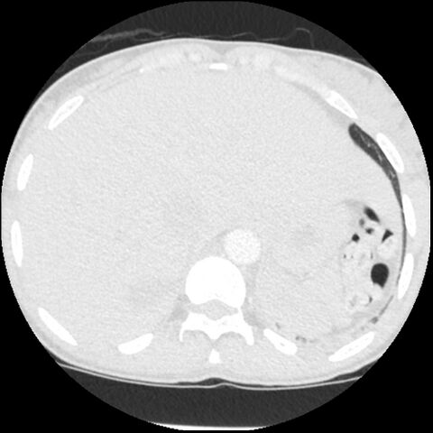
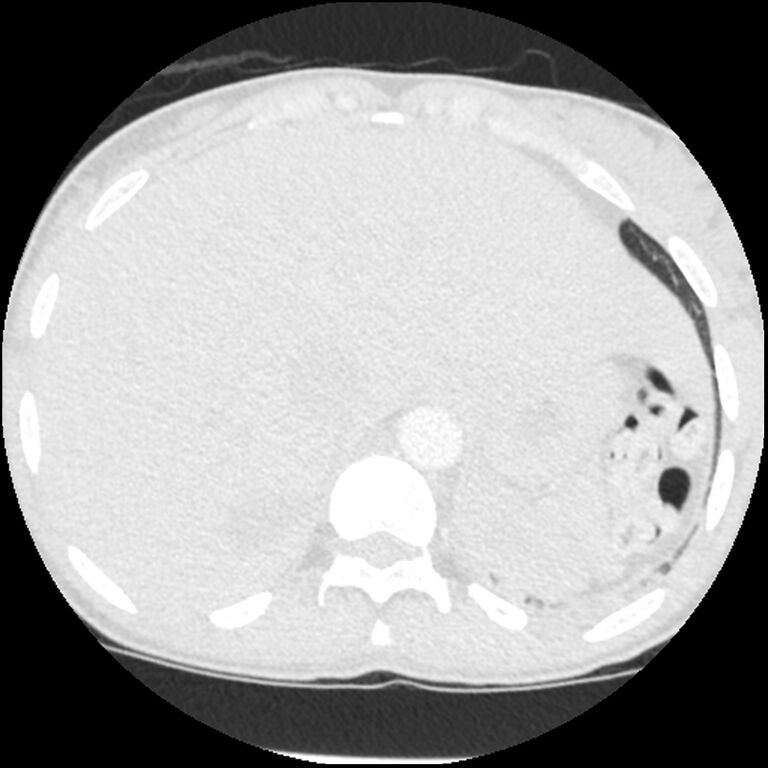
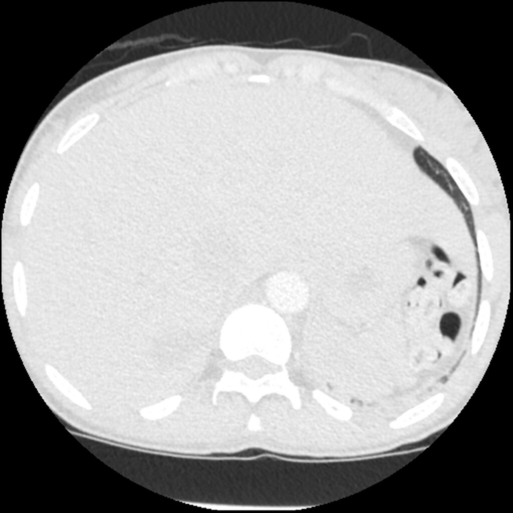
- Study findings: No acute pulmonary embolism. Mild cardiomegaly. Bilateral posterior lower lobe patchy consolidations, suggestive of either multifocal pneumonia or acute chest syndrome.
- Modality: CT
- System: Haematology
- Findings: Low lung volume with bilateral basal opacities.
- Published: 15th Aug 2017
- Source: https://radiopaedia.org/cases/acute-chest-syndrome-sickle-cell-disease-1
- Author: Hani Makky Al Salam
- Permission: http://creativecommons.org/licenses/by-nc-sa/3.0/
Licensing:
Attribution-NonCommercial-ShareAlike 3.0 Unported (CC BY-NC-SA 3.0)
File history
Click on a date/time to view the file as it appeared at that time.
| Date/Time | Thumbnail | Dimensions | User | Comment | |
|---|---|---|---|---|---|
| current | 22:33, 6 April 2021 | | 4,004 × 4,004 (2.45 MB) | Fæ (talk | contribs) | Radiopaedia project rID:42375 (batch #776-174 A174) |
You cannot overwrite this file.
File usage
The following page uses this file:
.jpg&oldid=80443)