File:Acute cholecystitis (Radiopaedia 70479-80598 Axial non-contrast 51).jpg
Jump to navigation
Jump to search
Size of this preview: 751 × 600 pixels. Other resolutions: 301 × 240 pixels | 601 × 480 pixels | 789 × 630 pixels.
Original file (789 × 630 pixels, file size: 233 KB, MIME type: image/jpeg)
Summary:
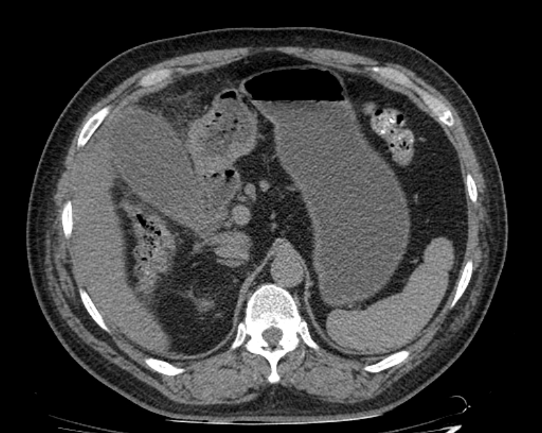
- Radiopaedia case ID: 70479
- Image ID: 51428033
- Image stack position: 51/170
- Plane projection: Axial
- Aux modality: non-contrast
- Modality: CT
- System: Hepatobiliary
- Findings: The gallbladder is distended, showing mild mural thickening and mural enhancement with surrounding inflammatory fat stranding. No radio-dense stone is detected in the lumen of the gallbladder and biliary ducts. No dilatation of the intra and extrahepatic biliary ducts is noted. Included sections from the lung base demonstrate; air space consolidatory changes with basal atelectasis predominantly in the right side. Incidental retro-aortic left renal vein. Rest of examination is unremarkable.
- Published: 21st Aug 2019
- Source: https://radiopaedia.org/cases/acute-cholecystitis-20
- Author: Farhad Farzam
- Permission: http://creativecommons.org/licenses/by-nc-sa/3.0/
Licensing:
Attribution-NonCommercial-ShareAlike 3.0 Unported (CC BY-NC-SA 3.0)
File history
Click on a date/time to view the file as it appeared at that time.
| Date/Time | Thumbnail | Dimensions | User | Comment | |
|---|---|---|---|---|---|
| current | 02:23, 7 April 2021 | | 789 × 630 (233 KB) | Fæ (talk | contribs) | Radiopaedia project rID:70479 (batch #778-51 A51) |
You cannot overwrite this file.
File usage
The following page uses this file:
.jpg&oldid=81483)