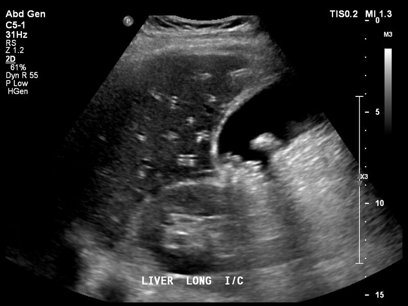
File:Acute cholecystitis (Radiopaedia 72392-82922 A 26).jpg
Jump to navigation
Jump to search

Size of this preview: 800 × 600 pixels. Other resolutions: 320 × 240 pixels | 640 × 480 pixels | 1,024 × 768 pixels.
Original file (1,024 × 768 pixels, file size: 93 KB, MIME type: image/jpeg)
Summary:
- Radiopaedia case ID: 72392
- Image ID: 51758315
- Image stack position: 26/40
- Study description: Abdomen
- Modality: Ultrasound
- System: Hepatobiliary
- Findings: The gallbladder is distended and edematous with surrounding hyperemia (wall thickness 0.4 cm). Mobile calculi and sludge are present within its lumen.Small volume free fluid in the right upper quadrant. The common bile duct is dilated to 0.8 cm with low-level internal echoes seen throughout its lumen to the level of the ampulla. No CBD stone identified anywhere along its course. There is intra-hepatic duct dilatation as seen on the recent CT. The pancreatic duct is not dilated. The main portal vein is patent and demonstrates normal directional blood flow. Conclusion:1. Features in keeping with acute calculus cholecystitis.2. Dilatation of the intra and extrahepatic ducts as seen on CT. The CBD was well visualized with sludge seen throughout its lumen. No evidence of a CBD stone.
- Published: 7th May 2020
- Source: https://radiopaedia.org/cases/acute-cholecystitis-26
- Author: Bruno Di Muzio
- Permission: http://creativecommons.org/licenses/by-nc-sa/3.0/
Licensing:
Attribution-NonCommercial-ShareAlike 3.0 Unported (CC BY-NC-SA 3.0)
File history
Click on a date/time to view the file as it appeared at that time.
| Date/Time | Thumbnail | Dimensions | User | Comment | |
|---|---|---|---|---|---|
| current | 04:59, 7 April 2021 |  | 1,024 × 768 (93 KB) | Fæ (talk | contribs) | Radiopaedia project rID:72392 (batch #780-26 A26) |
You cannot overwrite this file.
File usage
There are no pages that use this file.