File:Acute cholecystitis and incidental left sided IVC (Radiopaedia 49352-54459 Axial C+ portal venous phase 64).jpg
Jump to navigation
Jump to search
No higher resolution available.
Acute_cholecystitis_and_incidental_left_sided_IVC_(Radiopaedia_49352-54459_Axial_C+_portal_venous_phase_64).jpg (512 × 512 pixels, file size: 40 KB, MIME type: image/jpeg)
Summary:
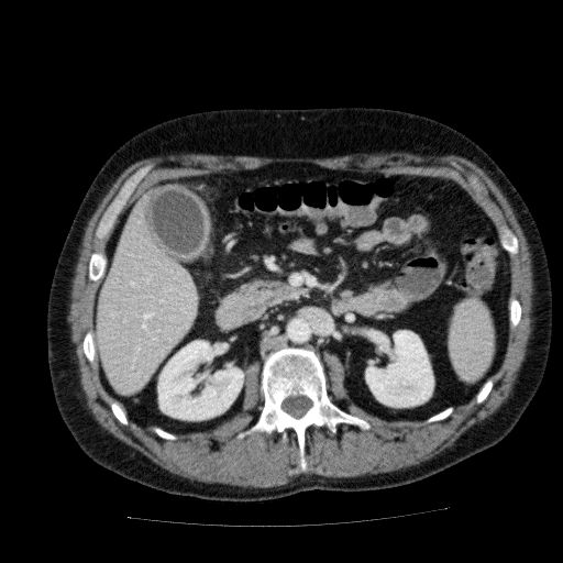
- Radiopaedia case ID: 49352
- Image ID: 26421092
- Image stack position: 64/177
- Plane projection: Axial
- Aux modality: C+ portal venous phase
- Modality: CT
- System: Hepatobiliary
- Findings: The gall bladder is thick-walled with abnormal enhancement of the wall and is surrounded by a thin rim of free fluid. There is no biliary dilatation or liver lesion. There is a left sided IVC but no other vascular anomaly or abnormality.Righ total hip replacement with old right iliac fracture. No other significant abnormality.
- Published: 13th May 2017
- Source: https://radiopaedia.org/cases/acute-cholecystitis-and-incidental-left-sided-ivc
- Author: Vikas Shah
- Permission: http://creativecommons.org/licenses/by-nc-sa/3.0/
Licensing:
Attribution-NonCommercial-ShareAlike 3.0 Unported (CC BY-NC-SA 3.0)
File history
Click on a date/time to view the file as it appeared at that time.
| Date/Time | Thumbnail | Dimensions | User | Comment | |
|---|---|---|---|---|---|
| current | 12:48, 7 April 2021 | | 512 × 512 (40 KB) | Fæ (talk | contribs) | Radiopaedia project rID:49352 (batch #793-64 A64) |
You cannot overwrite this file.
File usage
The following page uses this file:
.jpg&oldid=84952)