File:Acute cholecystitis complicated by pylephlebitis (Radiopaedia 65782-74915 Axial arterioportal phase T1 C+ fat sat 49).jpg
Jump to navigation
Jump to search
No higher resolution available.
Acute_cholecystitis_complicated_by_pylephlebitis_(Radiopaedia_65782-74915_Axial_arterioportal_phase_T1_C+_fat_sat_49).jpg (256 × 256 pixels, file size: 27 KB, MIME type: image/jpeg)
Summary:
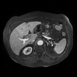
- Radiopaedia case ID: 65782
- Image ID: 45785830
- Image stack position: 49/91
- Plane projection: Axial arterioportal phase
- Aux modality: T1 C+ fat sat
- Modality: MRI
- System: Hepatobiliary
- Findings: The gallbladder demonstrates a thick and enhancing wall, containing numerous small gallstones. The intrahepatic biliary ducts are not dilated. The diameter of the common bile duct measures 7 mm with no distal stone. The common hepatic duct shows minimal stricture. The left portal vein and its branches are thrombosed. It appears dilated with high signal intensity on T1WI fat sat and T2WI, with no enhancement following IV contrast on arterioportal phase. The right portal vein is patent. An increased attenuation of the left hepatic parenchyma is noted on arterioportal phase usually due to increased arterial flow to compensate the decreased portal flow.
- Published: 23rd Jan 2019
- Source: https://radiopaedia.org/cases/acute-cholecystitis-complicated-by-pylephlebitis
- Author: Dr Ammar Haouimi
- Permission: http://creativecommons.org/licenses/by-nc-sa/3.0/
Licensing:
Attribution-NonCommercial-ShareAlike 3.0 Unported (CC BY-NC-SA 3.0)
File history
Click on a date/time to view the file as it appeared at that time.
| Date/Time | Thumbnail | Dimensions | User | Comment | |
|---|---|---|---|---|---|
| current | 17:56, 7 April 2021 | | 256 × 256 (27 KB) | Fæ (talk | contribs) | Radiopaedia project rID:65782 (batch #795-238 F49) |
You cannot overwrite this file.
File usage
The following page uses this file:
.jpg&oldid=86634)