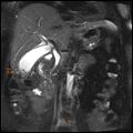
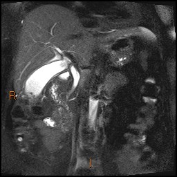
File:Acute cholecystitis with gallbladder neck calculus (Radiopaedia 42795-45971 Coronal T2 Half-fourier-acquired single-shot turbo spin echo (HASTE) 4).jpg
Jump to navigation
Jump to search
Acute_cholecystitis_with_gallbladder_neck_calculus_(Radiopaedia_42795-45971_Coronal_T2_Half-fourier-acquired_single-shot_turbo_spin_echo_(HASTE)_4).jpg (256 × 256 pixels, file size: 22 KB, MIME type: image/jpeg)
Summary:
- Radiopaedia case ID: 42795
- Image ID: 19364264
- Image stack position: 4/15
- Plane projection: Coronal
- Aux modality: T2 Half-fourier-acquired single-shot turbo spin echo (HASTE)
- Study findings: Thickened gallbladder, with a trace of pericholecystic fluid. Large impacted calculus at the gallbladder neck with multiple dependent calculus within the gallbladder.Mild intra and extrahepatic biliary dilatation with dilated CBD (8 mm). No ductal calculi or obstructing lesion identified. Low medial insertion of the cystic duct. Normal caliber pancreatic duct.
- Modality: MRI
- System: Hepatobiliary
- Findings: Thickwalled gallbladder (measuring 6mm) with large calculus obstructing the neck. Sludge in the gallbladder but no other calculi, nor in the ducts. Trace pericholecystic fluid. Dilated common bile duct (about 8mm) with intrahepatic biliary dilatation. Patent portal vein.
- Published: 9th Feb 2016
- Source: https://radiopaedia.org/cases/acute-cholecystitis-with-gallbladder-neck-calculus
- Author: Derek Smith
- Permission: http://creativecommons.org/licenses/by-nc-sa/3.0/
Licensing:
Attribution-NonCommercial-ShareAlike 3.0 Unported (CC BY-NC-SA 3.0)
File history
Click on a date/time to view the file as it appeared at that time.
| Date/Time | Thumbnail | Dimensions | User | Comment | |
|---|---|---|---|---|---|
| current | 19:50, 7 April 2021 |  | 256 × 256 (22 KB) | Fæ (talk | contribs) | Radiopaedia project rID:42795 (batch #798-4 A4) |
You cannot overwrite this file.
File usage
There are no pages that use this file.
