File:Acute complicated calculous cholecystitis (Radiopaedia 55202-61588 Image 1 1).png
Jump to navigation
Jump to search

Size of this preview: 800 × 379 pixels. Other resolutions: 320 × 152 pixels | 640 × 303 pixels | 1,318 × 624 pixels.
Original file (1,318 × 624 pixels, file size: 474 KB, MIME type: image/png)
Summary:
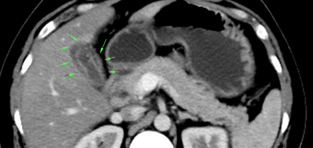
- Radiopaedia case ID: 55202
- Image ID: 32303781
- Plane projection: Image 1
- Study findings: Annotated images depicting the above findings. Image 1: Pericholecystic free fluidImage 2: Differential hepatic attenuationImage 3: Anterior segment right portal vein bland thrombusImage 4: Radiodense gall stone
- Modality: Annotated image
- System: Hepatobiliary
- Findings: Features of active gall bladder inflammation in the form of circumferential mural thickening, mural hyperenhancement, mild pericholecystic free fluid and dependent multiple <3 mm calculi. Filling defect seen in anterior branch of right portal vein. Secondary differential hepatic attenuation of segment V and VIII. Rest of the portal system, the hepatic arterial and venous systems are patent.
- Published: 25th Aug 2017
- Source: https://radiopaedia.org/cases/acute-complicated-calculous-cholecystitis
- Author: Varun Babu
- Permission: http://creativecommons.org/licenses/by-nc-sa/3.0/
Licensing:
Attribution-NonCommercial-ShareAlike 3.0 Unported (CC BY-NC-SA 3.0)
File history
Click on a date/time to view the file as it appeared at that time.
| Date/Time | Thumbnail | Dimensions | User | Comment | |
|---|---|---|---|---|---|
| current | 04:40, 8 April 2021 | | 1,318 × 624 (474 KB) | Fæ (talk | contribs) | Radiopaedia project rID:55202 (batch #802-1 A1) |
You cannot overwrite this file.
File usage
There are no pages that use this file.
.png&oldid=90363)