File:Acute disseminated encephalomyelitis (ADEM) (Radiopaedia 58877-66115 Axial T1 C+ 19).jpg
Jump to navigation
Jump to search
No higher resolution available.
Acute_disseminated_encephalomyelitis_(ADEM)_(Radiopaedia_58877-66115_Axial_T1_C+_19).jpg (512 × 512 pixels, file size: 86 KB, MIME type: image/jpeg)
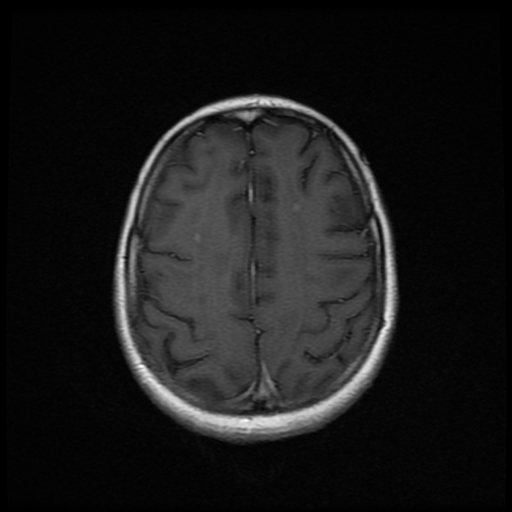
Summary:
- Radiopaedia case ID: 58877
- Image ID: 36764754
- Image stack position: 19/24
- Plane projection: Axial
- Aux modality: T1 C+
- Modality: MRI
- System: Central Nervous System
- Findings: Abnormal confluent regions of T2 and FLAIR high signal, situated in a sub-cortical location. Post contrast series reveal multiple scattered punctuate like enhancing lesions range in size from 4 mm to 8 mm. Abnormal high signal is demonstrated within both ADC map (not shown) and DWI denotes T2 shine through effect. No diffusion restriction. No involvement of the gray matter.
- Published: 13th Mar 2018
- Source: https://radiopaedia.org/cases/acute-disseminated-encephalomyelitis-adem-4
- Author: Heba Abdelmonem
- Permission: http://creativecommons.org/licenses/by-nc-sa/3.0/
Licensing:
Attribution-NonCommercial-ShareAlike 3.0 Unported (CC BY-NC-SA 3.0)
File history
Click on a date/time to view the file as it appeared at that time.
| Date/Time | Thumbnail | Dimensions | User | Comment | |
|---|---|---|---|---|---|
| current | 11:31, 9 April 2021 | | 512 × 512 (86 KB) | Fæ (talk | contribs) | Radiopaedia project rID:58877 (batch #818-91 D19) |
You cannot overwrite this file.
File usage
There are no pages that use this file.
_(Radiopaedia_58877-66115_Axial_T1_C%2B_19).jpg&oldid=98710)