File:Acute disseminated encephalomyelitis (Radiopaedia 37253-39033 Axial DWI 50).jpg
Jump to navigation
Jump to search
No higher resolution available.
Acute_disseminated_encephalomyelitis_(Radiopaedia_37253-39033_Axial_DWI_50).jpg (594 × 594 pixels, file size: 64 KB, MIME type: image/jpeg)
Summary:
- Radiopaedia case ID: 37253
- Image ID: 13228281
- Image stack position: 50/54
- Plane projection: Axial
- Aux modality: DWI
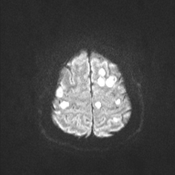
- Study findings: Huge number of well delineated high T2/FLAIR, non restricting enhancing lesions of variable size throughout both cerebral hemispheres in the deep white matter, juxta-cortical regions and left thalamus. No perilesional edema. Normal posterior fossa.
- Modality: MRI
- System: Central Nervous System
- Findings: Extensive ill-defined foci of low attenutation in throughout both cerebral hemispheres in the deep white matter, juxta-cortical regions and left thalamus. Normal posterior fossa.
- Published: 24th Jun 2015
- Source: https://radiopaedia.org/cases/acute-disseminated-encephalomyelitis-6
- Author: Ian Bickle
- Permission: http://creativecommons.org/licenses/by-nc-sa/3.0/
Licensing:
Attribution-NonCommercial-ShareAlike 3.0 Unported (CC BY-NC-SA 3.0)
File history
Click on a date/time to view the file as it appeared at that time.
| Date/Time | Thumbnail | Dimensions | User | Comment | |
|---|---|---|---|---|---|
| current | 20:08, 8 April 2021 | | 594 × 594 (64 KB) | Fæ (talk | contribs) | Radiopaedia project rID:37253 (batch #810-130 D50) |
You cannot overwrite this file.
File usage
The following page uses this file:
.jpg&oldid=93536)