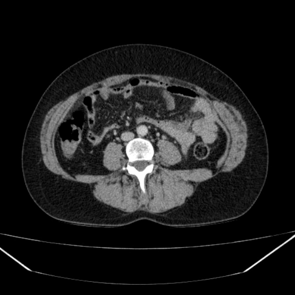
File:Acute diverticulitis (Radiopaedia 34183-35442 Axial C+ portal venous phase 80).jpg
Jump to navigation
Jump to search
Size of this preview: 600 × 600 pixels. Other resolutions: 240 × 240 pixels | 607 × 607 pixels.
Original file (607 × 607 pixels, file size: 81 KB, MIME type: image/jpeg)
Summary:
- Radiopaedia case ID: 34183
- Image ID: 10936579
- Image stack position: 80/152
- Plane projection: Axial
- Aux modality: C+ portal venous phase
- Study description: CT abdomen and pelvis
- Modality: CT
- System: Gastrointestinal
- Findings: The colon has multiple diverticula in the sigmoid and descending portions, with changes in attenuation of the adjacent fat, without any collections or extraluminal air. Liver size is normal without focal lesions. Pancreas, spleen, and adrenal glands are normal. The left kidney has multiple areas of parenchymal thinning and is decreased in size. Small duodenal diverticulum.
- Published: 11th Feb 2015
- Source: https://radiopaedia.org/cases/acute-diverticulitis-3
- Author: David Cuete
- Permission: http://creativecommons.org/licenses/by-nc-sa/3.0/
Licensing:
Attribution-NonCommercial-ShareAlike 3.0 Unported (CC BY-NC-SA 3.0)
File history
Click on a date/time to view the file as it appeared at that time.
| Date/Time | Thumbnail | Dimensions | User | Comment | |
|---|---|---|---|---|---|
| current | 18:14, 9 April 2021 | | 607 × 607 (81 KB) | Fæ (talk | contribs) | Radiopaedia project rID:34183 (batch #823-171 B80) |
You cannot overwrite this file.
File usage
The following page uses this file:
.jpg&oldid=101013)