File:Acute gangrenous cholecystitis (Radiopaedia 10123-10661 Coronal C+ portal venous phase 9).jpg
Jump to navigation
Jump to search

Size of this preview: 600 × 600 pixels. Other resolutions: 240 × 240 pixels | 480 × 480 pixels | 792 × 792 pixels.
Original file (792 × 792 pixels, file size: 272 KB, MIME type: image/jpeg)
Summary:
- Radiopaedia case ID: 10123
- Image ID: 495725
- Image stack position: 9/32
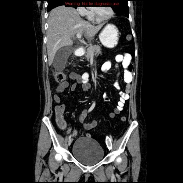
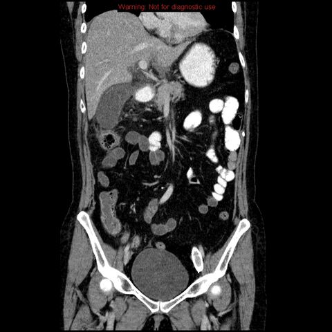
- Plane projection: Coronal
- Aux modality: C+ portal venous phase
- Modality: CT
- System: Hepatobiliary
- Findings: The gallbladder is distended and contains some high-density material. The wall is of variable thickness and density with patchy enhancement. There is pericholecystic fluid and inflammatory change with peritoneal and retroperitoneal fluid, and a right-sided pleural effusion. There are a couple of gallstones, but nothing obstructive in the CBD or head of the pancreas. Gangrenous cholecystitis is considered the most likely diagnosis because of localized right upper quadrant inflammatory change and patchy gallbladder wall enhancement.
- Published: 1st Jul 2010
- Source: https://radiopaedia.org/cases/acute-gangrenous-cholecystitis
- Author: Jeremy Jones
- Permission: http://creativecommons.org/licenses/by-nc-sa/3.0/
Licensing:
Attribution-NonCommercial-ShareAlike 3.0 Unported (CC BY-NC-SA 3.0)
File history
Click on a date/time to view the file as it appeared at that time.
| Date/Time | Thumbnail | Dimensions | User | Comment | |
|---|---|---|---|---|---|
| current | 19:48, 10 April 2021 | | 792 × 792 (272 KB) | Fæ (talk | contribs) | Radiopaedia project rID:10123 (batch #846-122 B9) |
You cannot overwrite this file.
File usage
There are no pages that use this file.
.jpg&oldid=109539)