File:Acute hemorrhage in a renal cyst in polycystic disease (MRI) (Radiopaedia 41702-44642 Coronal T2 fat sat 1).jpg
Jump to navigation
Jump to search
Acute_hemorrhage_in_a_renal_cyst_in_polycystic_disease_(MRI)_(Radiopaedia_41702-44642_Coronal_T2_fat_sat_1).jpg (512 × 512 pixels, file size: 113 KB, MIME type: image/jpeg)
Summary:
- Radiopaedia case ID: 41702
- Image ID: 18168378
- Plane projection: Coronal
- Aux modality: T2 fat sat
- Study description: MRI to assist in interpretation of CT findings
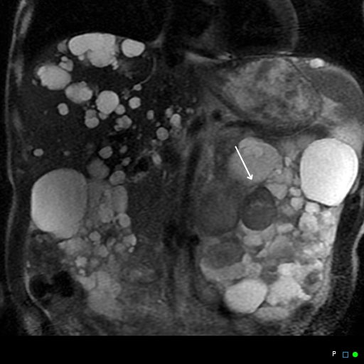
- Study findings: Confirmed blood in antero-medial left kidney based on morphology and signal characteristics on T1 and T2 as well as lack of enhancement.
- Modality: MRI
- System: Urogenital
- Findings: Typical findings of polycystic disease with enlarged cystic kidneys. Note multiple hyperdense cysts on the left including the antero-medial kidney (arrow), suspicious of hemorrhage.
- Published: 16th Dec 2015
- Source: https://radiopaedia.org/cases/acute-haemorrhage-in-a-renal-cyst-in-polycystic-disease-mri
- Author: Chris O'Donnell
- Permission: http://creativecommons.org/licenses/by-nc-sa/3.0/
Licensing:
Attribution-NonCommercial-ShareAlike 3.0 Unported (CC BY-NC-SA 3.0)
File history
Click on a date/time to view the file as it appeared at that time.
| Date/Time | Thumbnail | Dimensions | User | Comment | |
|---|---|---|---|---|---|
| current | 21:39, 10 April 2021 |  | 512 × 512 (113 KB) | Fæ (talk | contribs) | Radiopaedia project rID:41702 (batch #849-1 A1) |
You cannot overwrite this file.
File usage
There are no pages that use this file.
