File:Acute hemorrhage into adrenal metastasis from NSCLC (Radiopaedia 43135-46429 Axial C+ arterial phase 2).jpg
Jump to navigation
Jump to search

Size of this preview: 599 × 600 pixels. Other resolutions: 240 × 240 pixels | 479 × 480 pixels | 707 × 708 pixels.
Original file (707 × 708 pixels, file size: 147 KB, MIME type: image/jpeg)
Summary:
- Radiopaedia case ID: 43135
- Image ID: 19780949
- Image stack position: 2/13
- Plane projection: Axial
- Aux modality: C+ arterial phase
- Modality: CT
- System: Urogenital
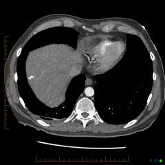
- Findings: Pre-chemotherapy CT performed 10 days earlier shown in series 2 and 4. Irregular mass in the right lung apex (arrow) is associated with a large right adrenal mass lesion with central low density indicative of necrosis (arrow). Right lung base is clear. Subsequent CTPA shows marked right lung atelectasis with pleural fluid with a dramatic increase in size of the metastasis with central high denisty indicative of hemorrhage (arrow). This explains the right sided pleuritic symptoms and hypotension. No signs of PE.
- Published: 25th Feb 2016
- Source: https://radiopaedia.org/cases/acute-haemorrhage-into-adrenal-metastasis-from-nsclc
- Author: Chris O'Donnell
- Permission: http://creativecommons.org/licenses/by-nc-sa/3.0/
Licensing:
Attribution-NonCommercial-ShareAlike 3.0 Unported (CC BY-NC-SA 3.0)
File history
Click on a date/time to view the file as it appeared at that time.
| Date/Time | Thumbnail | Dimensions | User | Comment | |
|---|---|---|---|---|---|
| current | 21:50, 10 April 2021 | | 707 × 708 (147 KB) | Fæ (talk | contribs) | Radiopaedia project rID:43135 (batch #850-21 B2) |
You cannot overwrite this file.
File usage
There are no pages that use this file.
.jpg&oldid=110198)