File:Acute hemorrhagic leukoencephalitis (Radiopaedia 14076-43697 Axial non-contrast 9).jpg
Jump to navigation
Jump to search
No higher resolution available.
Acute_hemorrhagic_leukoencephalitis_(Radiopaedia_14076-43697_Axial_non-contrast_9).jpg (512 × 512 pixels, file size: 115 KB, MIME type: image/jpeg)
Summary:
- Radiopaedia case ID: 14076
- Image ID: 1119999
- Image stack position: 9/51
- Plane projection: Axial
- Aux modality: non-contrast
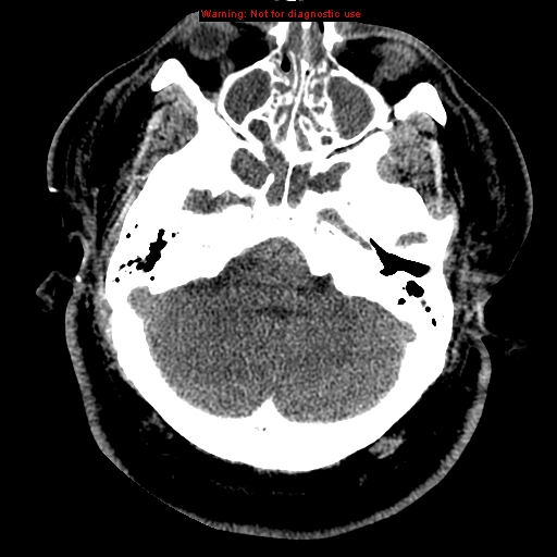
- Study findings: Urgent decompressive bilateral fronto-temporal craniectomies were performed. CT Brain 9 days following the initial presentation shows established bilateral posterior cerebral artery territory infarcts as a result of the previous descending transtentorial herniation.
- Modality: CT
- System: Central Nervous System
- Findings: Initial CT Brain demonstrates diffuse cerebral white matter hypoattenuation including involvement of the corpus callosum. There is associated generalized cerebral sulcal effacement. Brain biopsy was performed the following day showing acute hemorrhagic leukoencephalitis (see path report below).
- Published: 21st Jun 2011
- Source: https://radiopaedia.org/cases/acute-haemorrhagic-leukoencephalitis
- Author: Andrew Dixon
- Permission: http://creativecommons.org/licenses/by-nc-sa/3.0/
Licensing:
Attribution-NonCommercial-ShareAlike 3.0 Unported (CC BY-NC-SA 3.0)
File history
Click on a date/time to view the file as it appeared at that time.
| Date/Time | Thumbnail | Dimensions | User | Comment | |
|---|---|---|---|---|---|
| current | 22:34, 10 April 2021 | | 512 × 512 (115 KB) | Fæ (talk | contribs) | Radiopaedia project rID:14076 (batch #851-9 A9) |
You cannot overwrite this file.
File usage
The following page uses this file:
.jpg&oldid=110451)