File:Acute hepatitis (Radiopaedia 51456-57211 C 84).jpg
Jump to navigation
Jump to search

Size of this preview: 489 × 600 pixels. Other resolutions: 196 × 240 pixels | 391 × 480 pixels | 626 × 768 pixels | 835 × 1,024 pixels | 1,504 × 1,844 pixels.
Original file (1,504 × 1,844 pixels, file size: 86 KB, MIME type: image/jpeg)
Summary:
- Radiopaedia case ID: 51456
- Image ID: 28804020
- Image stack position: 84/90
- Modality: MRI
- System: Gastrointestinal
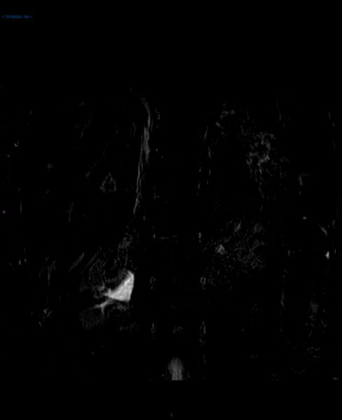
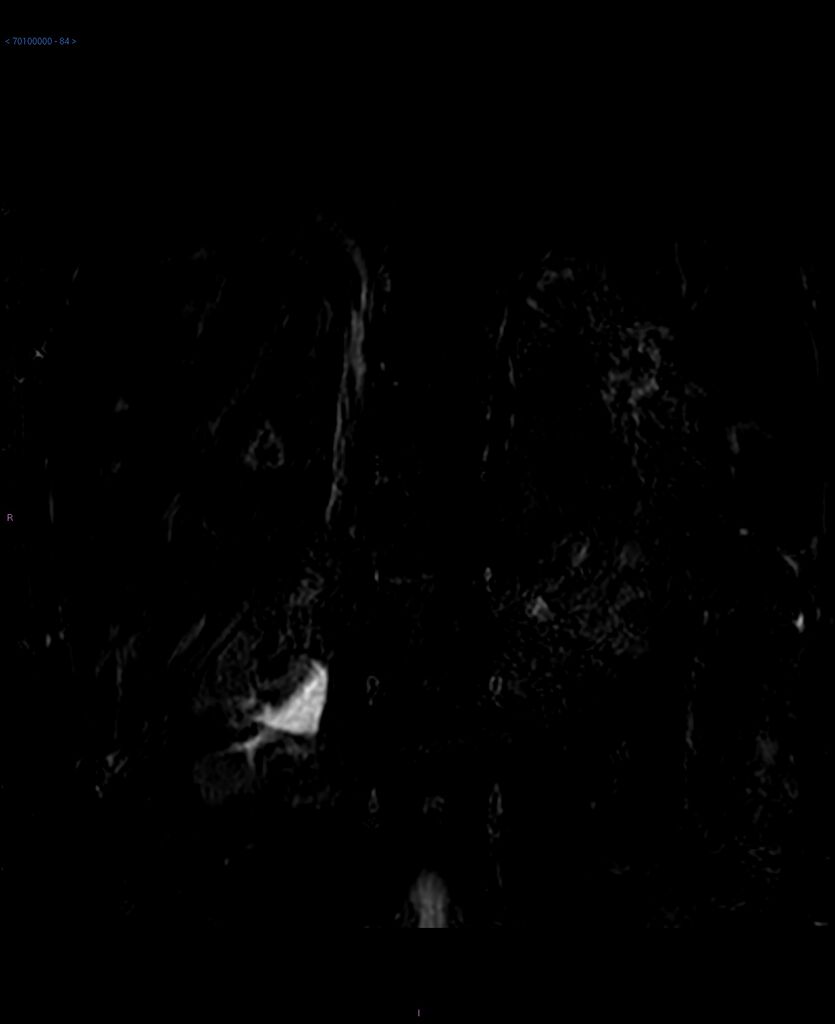
- Findings: There is severe gallbladder wall T2 hyperintensity in keeping with edema, which compresses the mucosa of the gallbladder. Cystic duct, CBD pancreatic duct are within normal limits with no obstructing lesions seen.Marked high T2 signal is also demonstrated within the periportal spaces consistent with edema. Liver also appears slightly enlarged with recanalization of the umbilical vein noted. Overall findings suggestive of acute hepatitis and could account for the markedly deranged LFTs.
- Published: 16th Feb 2017
- Source: https://radiopaedia.org/cases/acute-hepatitis-4
- Author: Mark Hall
- Permission: http://creativecommons.org/licenses/by-nc-sa/3.0/
Licensing:
Attribution-NonCommercial-ShareAlike 3.0 Unported (CC BY-NC-SA 3.0)
File history
Click on a date/time to view the file as it appeared at that time.
| Date/Time | Thumbnail | Dimensions | User | Comment | |
|---|---|---|---|---|---|
| current | 02:34, 11 April 2021 | | 1,504 × 1,844 (86 KB) | Fæ (talk | contribs) | Radiopaedia project rID:51456 (batch #855-162 C84) |
You cannot overwrite this file.
File usage
The following page uses this file:
.jpg&oldid=111811)