File:Acute hippocampal infarction (Radiopaedia 10610-11072 B 1).jpg
Jump to navigation
Jump to search
No higher resolution available.
Acute_hippocampal_infarction_(Radiopaedia_10610-11072_B_1).jpg (512 × 512 pixels, file size: 94 KB, MIME type: image/jpeg)
Summary:
- Radiopaedia case ID: 10610
- Image ID: 538574
- Image stack position: 1/3
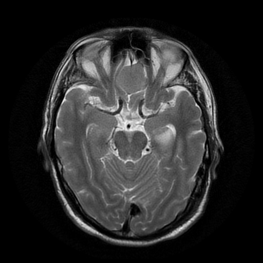
- Aux modality: T2
- Study findings: On subsequent MRI, there is swelling of the left hippocampus with T2 and DWI hyperintensity involving the hippocampal body and tail, consistent with acute hippocampal infarction. There is also a meningioma arising from the planum sphenoidale.
- Modality: MRI
- System: Central Nervous System
- Findings: CT demonstrates hypoattenuation in the region of the left hippocampus in keeping with an acute hippocampal infarction. Frontal meningioma.
- Published: 25th Aug 2010
- Source: https://radiopaedia.org/cases/acute-hippocampal-infarction
- Author: Alexandra Stanislavsky
- Permission: http://creativecommons.org/licenses/by-nc-sa/3.0/
Licensing:
Attribution-NonCommercial-ShareAlike 3.0 Unported (CC BY-NC-SA 3.0)
File history
Click on a date/time to view the file as it appeared at that time.
| Date/Time | Thumbnail | Dimensions | User | Comment | |
|---|---|---|---|---|---|
| current | 05:53, 11 April 2021 | | 512 × 512 (94 KB) | Fæ (talk | contribs) | Radiopaedia project rID:10610 (batch #867-2 B1) |
You cannot overwrite this file.
File usage
There are no pages that use this file.
.jpg&oldid=112911)