File:Acute internal carotid artery dissection (Radiopaedia 53541-59630 Axial C+ arterial phase 63).jpg
Jump to navigation
Jump to search
No higher resolution available.
Acute_internal_carotid_artery_dissection_(Radiopaedia_53541-59630_Axial_C+_arterial_phase_63).jpg (420 × 435 pixels, file size: 8 KB, MIME type: image/jpeg)
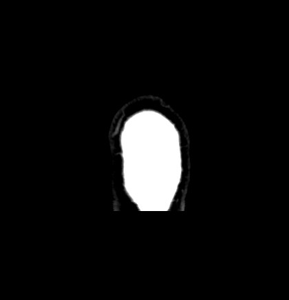
Summary:
- Radiopaedia case ID: 53541
- Image ID: 30379693
- Image stack position: 63/64
- Plane projection: Axial
- Aux modality: C+ arterial phase
- Study findings: CT angiogram of the brain. Left internal carotid artery dissection is better appreciated in this study. The left internal carotid artery demonstrates an abnormal vessel contour, with mildly narrowed distal segment surrounded by a non-enhancing crescent-shaped mural thrombus. The rest of the circle of Willis demonstrates normal enhancement. No other abnormality is seen.
- Modality: CT
- System: Central Nervous System
- Findings: CT brain study. No evidence of sulcal effacement, acute hemorrhage or ischemic changes within the brain. Ventricular pattern is appropriate for age. No acute fractures seen. The cervical segment of the left internal carotid artery demonstrates a narrowed lumen with a crescent-shaped hyperattenuating focus, favored to be a mural thrombus that extends towards the skull base. This is suspicious for acute left internal carotid artery dissection, especially given the presenting history.
- Published: 12th Sep 2017
- Source: https://radiopaedia.org/cases/acute-internal-carotid-artery-dissection
- Author: Andrew Dixon
- Permission: http://creativecommons.org/licenses/by-nc-sa/3.0/
Licensing:
Attribution-NonCommercial-ShareAlike 3.0 Unported (CC BY-NC-SA 3.0)
File history
Click on a date/time to view the file as it appeared at that time.
| Date/Time | Thumbnail | Dimensions | User | Comment | |
|---|---|---|---|---|---|
| current | 16:59, 11 April 2021 | | 420 × 435 (8 KB) | Fæ (talk | contribs) | Radiopaedia project rID:53541 (batch #879-63 A63) |
You cannot overwrite this file.
File usage
The following page uses this file:
.jpg&oldid=116174)