File:Acute lupus nephritis (Radiopaedia 45832-50108 B 1).jpg
Jump to navigation
Jump to search
Size of this preview: 800 × 546 pixels. Other resolutions: 320 × 218 pixels | 640 × 437 pixels | 1,024 × 699 pixels | 1,280 × 873 pixels | 2,560 × 1,747 pixels | 4,847 × 3,307 pixels.
Original file (4,847 × 3,307 pixels, file size: 2.06 MB, MIME type: image/jpeg)
Summary:
- Radiopaedia case ID: 45832
- Image ID: 23251320
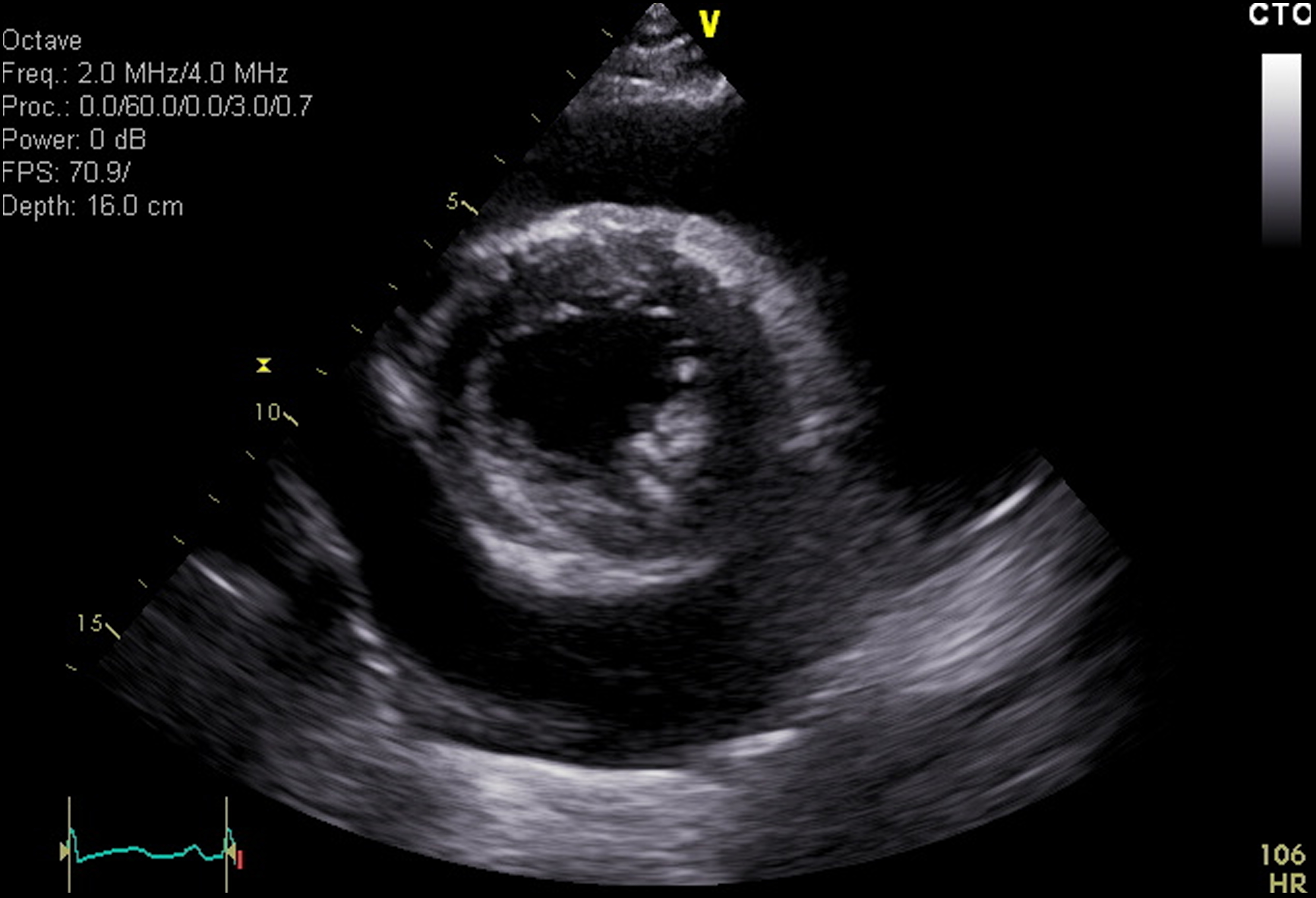
- Plane projection: Transverse
- Study description: Transthoracic Echocardiogram
- Study findings: There is a large circumferential pericardial effusion with signs suggestive of early tamponade. Structurally normal aortic, mitral, tricuspid and pulmonic valves with trace tricuspid and pulmonic regurgitation. Normal chamber dimensions with normal right and left ventricular systolic function, estimated LV EF is 60-65%.
- Modality: Ultrasound
- System: Urogenital
- Findings: Flask-shaped enlargement of the cardiac silhouette in keeping with pericardial effusion. The lungs are clear.
- Published: 28th Oct 2017
- Source: https://radiopaedia.org/cases/acute-lupus-nephritis
- Author: Hani Makky Al Salam
- Permission: http://creativecommons.org/licenses/by-nc-sa/3.0/
Licensing:
Attribution-NonCommercial-ShareAlike 3.0 Unported (CC BY-NC-SA 3.0)
File history
Click on a date/time to view the file as it appeared at that time.
| Date/Time | Thumbnail | Dimensions | User | Comment | |
|---|---|---|---|---|---|
| current | 06:34, 13 April 2021 | | 4,847 × 3,307 (2.06 MB) | Fæ (talk | contribs) | Radiopaedia project rID:45832 (batch #902-2 B1) |
You cannot overwrite this file.
File usage
There are no pages that use this file.
.jpg&oldid=127716)