File:Acute lymphoblastic leukemia (Radiopaedia 49049-54145 Coronal PD fat sat 1).jpg
Jump to navigation
Jump to search

Size of this preview: 600 × 600 pixels. Other resolutions: 240 × 240 pixels | 480 × 480 pixels | 768 × 768 pixels | 1,024 × 1,024 pixels | 1,474 × 1,474 pixels.
Original file (1,474 × 1,474 pixels, file size: 219 KB, MIME type: image/jpeg)
Summary:
- Radiopaedia case ID: 49049
- Image ID: 29166125
- Image stack position: 1/23
- Plane projection: Coronal
- Aux modality: PD fat sat
- Study description: Knee MRI
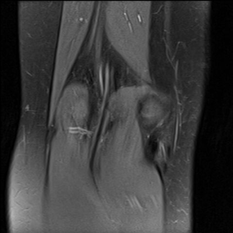
- Study findings: Numerous well circumscribed intramedullary lesions of variable size are demonstrated throughout the imaged periarticular femur, tibia, fibula and patella, involving both the metadiaphyses and epiphyses. The lesions are of low T1 signal. Most are of high signal on the PD fat sat (fluid sensitive) sequences, and low signal on the PD acquisition. There is minimal cortical scalloping, but no overt lytic process. Additionally, there is more confluent fluid signal within the anterior tibial metaphysis, with suspected anteromedial microtrabecular fracture. The cruciate and collateral ligaments, menisci and chondral surfaces are intact.
- Modality: MRI
- System: Haematology
- Findings: Subtle, patchy sclerotic changes in the distal femoral and proximal tibial metaphyses.
- Published: 15th Jan 2018
- Source: https://radiopaedia.org/cases/acute-lymphoblastic-leukaemia-1
- Author: Alexandra Stanislavsky
- Permission: http://creativecommons.org/licenses/by-nc-sa/3.0/
Licensing:
Attribution-NonCommercial-ShareAlike 3.0 Unported (CC BY-NC-SA 3.0)
File history
Click on a date/time to view the file as it appeared at that time.
| Date/Time | Thumbnail | Dimensions | User | Comment | |
|---|---|---|---|---|---|
| current | 10:17, 13 April 2021 | | 1,474 × 1,474 (219 KB) | Fæ (talk | contribs) | Radiopaedia project rID:49049 (batch #904-1 A1) |
You cannot overwrite this file.
File usage
There are no pages that use this file.
.jpg&oldid=128846)