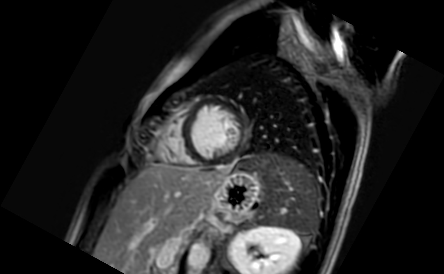
File:Acute myocarditis (Radiopaedia 77023-88967 Short axis stack LGE 5).jpg
Jump to navigation
Jump to search
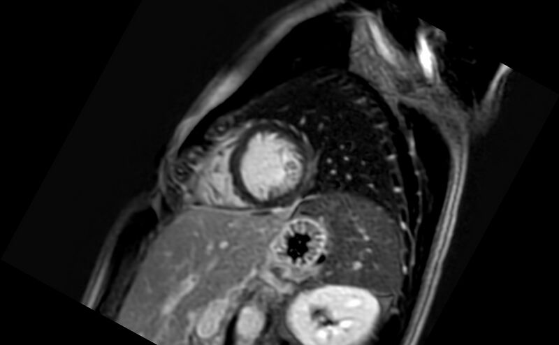
Size of this preview: 800 × 494 pixels. Other resolutions: 320 × 198 pixels | 640 × 395 pixels | 1,024 × 632 pixels | 1,526 × 942 pixels.
Original file (1,526 × 942 pixels, file size: 130 KB, MIME type: image/jpeg)
Summary:
| Description |
|
| Date | Published: 5th May 2020 |
| Source | https://radiopaedia.org/cases/acute-myocarditis-3 |
| Author | Tamara Razon Cuenza |
| Permission (Permission-reusing-text) |
http://creativecommons.org/licenses/by-nc-sa/3.0/ |
Licensing:
Attribution-NonCommercial-ShareAlike 3.0 Unported (CC BY-NC-SA 3.0)
File history
Click on a date/time to view the file as it appeared at that time.
| Date/Time | Thumbnail | Dimensions | User | Comment | |
|---|---|---|---|---|---|
| current | 22:22, 14 April 2021 | | 1,526 × 942 (130 KB) | Fæ (talk | contribs) | Radiopaedia project rID:77023 (batch #938-50 E5) |
You cannot overwrite this file.
File usage
There are no pages that use this file.
.jpg&oldid=140193)