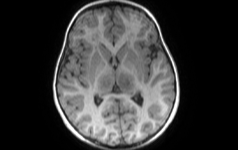
File:Acute necrotizing encephalitis of childhood (Radiopaedia 67356-76737 Axial T1 46).jpg
Jump to navigation
Jump to search
Acute_necrotizing_encephalitis_of_childhood_(Radiopaedia_67356-76737_Axial_T1_46).jpg (783 × 494 pixels, file size: 83 KB, MIME type: image/jpeg)
Summary:
| Description |
|
| Date | Published: 2nd Apr 2019 |
| Source | https://radiopaedia.org/cases/acute-necrotizing-encephalitis-of-childhood-1 |
| Author | Ahmed Abdrabou |
| Permission (Permission-reusing-text) |
http://creativecommons.org/licenses/by-nc-sa/3.0/ |
Licensing:
Attribution-NonCommercial-ShareAlike 3.0 Unported (CC BY-NC-SA 3.0)
File history
Click on a date/time to view the file as it appeared at that time.
| Date/Time | Thumbnail | Dimensions | User | Comment | |
|---|---|---|---|---|---|
| current | 13:51, 15 April 2021 |  | 783 × 494 (83 KB) | Fæ (talk | contribs) | Radiopaedia project rID:67356 (batch #948-46 A46) |
You cannot overwrite this file.
File usage
The following page uses this file:
