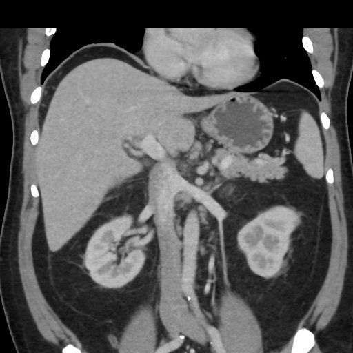
File:Acute on chronic calculous cholecystitis (Radiopaedia 55562-62050 Coronal C+ portal venous phase 36).jpg
Jump to navigation
Jump to search
Acute_on_chronic_calculous_cholecystitis_(Radiopaedia_55562-62050_Coronal_C+_portal_venous_phase_36).jpg (512 × 512 pixels, file size: 70 KB, MIME type: image/jpeg)
Summary:
| Description |
|
| Date | Published: 15th Sep 2017 |
| Source | https://radiopaedia.org/cases/acute-on-chronic-calculous-cholecystitis |
| Author | Varun Babu |
| Permission (Permission-reusing-text) |
http://creativecommons.org/licenses/by-nc-sa/3.0/ |
Licensing:
Attribution-NonCommercial-ShareAlike 3.0 Unported (CC BY-NC-SA 3.0)
File history
Click on a date/time to view the file as it appeared at that time.
| Date/Time | Thumbnail | Dimensions | User | Comment | |
|---|---|---|---|---|---|
| current | 17:18, 15 April 2021 |  | 512 × 512 (70 KB) | Fæ (talk | contribs) | Radiopaedia project rID:55562 (batch #951-77 B36) |
You cannot overwrite this file.
File usage
There are no pages that use this file.
