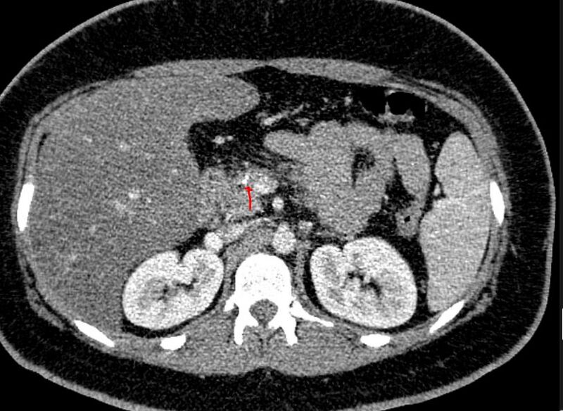
File:Acute on chronic pancreatitis (Radiopaedia 80902-94538 B 1).JPG
Jump to navigation
Jump to search

Size of this preview: 800 × 584 pixels. Other resolutions: 320 × 234 pixels | 640 × 467 pixels | 814 × 594 pixels.
Original file (814 × 594 pixels, file size: 103 KB, MIME type: image/jpeg)
Summary:
| Description |
|
| Date | Published: 18th Aug 2020 |
| Source | https://radiopaedia.org/cases/acute-on-chronic-pancreatitis-2 |
| Author | Faeze Salahshour |
| Permission (Permission-reusing-text) |
http://creativecommons.org/licenses/by-nc-sa/3.0/ |
Licensing:
Attribution-NonCommercial-ShareAlike 3.0 Unported (CC BY-NC-SA 3.0)
File history
Click on a date/time to view the file as it appeared at that time.
| Date/Time | Thumbnail | Dimensions | User | Comment | |
|---|---|---|---|---|---|
| current | 21:39, 15 April 2021 |  | 814 × 594 (103 KB) | Fæ (talk | contribs) | Radiopaedia project rID:80902 (batch #955-2 B1) |
You cannot overwrite this file.
File usage
There are no pages that use this file.