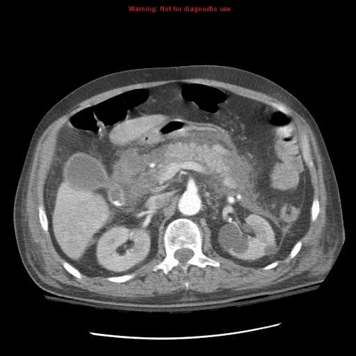
File:Acute pancreatitis with incidental pancreatic lipoma (Radiopaedia 10190-10730 Axial C+ arterial phase 38).jpg

From NC Commons
Jump to navigation Jump to search

Summary:

Description
  • Radiopaedia case ID: 10190
  • Image ID: 519030
  • Image stack position: 38/64
  • Plane projection: Axial
  • Aux modality: C+ arterial phase
  • Modality: CT
  • System: Hepatobiliary
  • Findings: The pancreas is swollen, associated with extensive peripancreatic stranding and fluid in the lesser sac, extending to the pararenal spaces, more on the left side, with minimal perisplenic and perihepatic free fluid. A very small part of the head of the pancreas appears to be less enhancing compared to the rest of the gland suspicious for necrosis. No gas locule can be seen in the pancreas. No walled off collection or pseudocysts can be seen. There is a small hypodense, well-defined, non-enhancing lesion seen in the tail of the pancreas. Gallbladder contains radiopaque stones in the neck, with no CT features of acute cholecystitis. The celiac and SMA are patent. No splenic artery pseudoaneurysm. Portal, splenic and superior mesenteric veins are patent, with no evidence of thrombosis. Left sided pleural effusion (minimal) and associated basal collapse/consolidation. Interpretation: Features of acute pancreatitis, likely due to cholelithiasis. A small hypoenhancing pancreatic head portion is suggestive of necrosis. Small, non-enhancing hypodense pancreatic tail lesion could represent a small cyst (pseudocyst is though less likely given the timing of the CT since the symptoms) or other lesions, such as lipoma, but needs further evaluation once the patient clinically improves.
Date Published: 20th Jul 2010
Source https://radiopaedia.org/cases/acute-pancreatitis-with-incidental-pancreatic-lipoma
Author Hani Makky Al Salam
Permission
(Permission-reusing-text)
http://creativecommons.org/licenses/by-nc-sa/3.0/

Licensing:

Attribution-NonCommercial-ShareAlike 3.0 Unported (CC BY-NC-SA 3.0)

File history

Click on a date/time to view the file as it appeared at that time.

Date/TimeThumbnailDimensionsUserComment
current20:50, 17 April 2021Thumbnail for version as of 20:50, 17 April 2021512 × 512 (112 KB) (talk | contribs)Radiopaedia project rID:10190 (batch #1018-38 A38)